Clinical Approach to a Case of Uveitis
A simplified way to decode a case of uveitis for the ophthalmologists
CLASSIFICATION OF UVEITIS
Proper classification of uveitic entities is essential to avoid confusion and misinterpretation among ophthalmologists. There have been many classifications proposed for uveitis. This section deals with the various available classification systems of uveitis.
Clinicopathological or Woods Classification:
One of the oldest classifications of uveitis is Woods classification (also known as pathological or clinicopathological classification). This was proposed by Alan Churchill Woods. Uveitis is classified as granulomatous or nongranulomatous on the basis of the predominant clinical characteristics.
|
Granulomatous Uveitis |
Nongranulomatous Uveitis |
|
Insidious onset and chronic course |
Sudden onset and acute course |
|
Absent or mild congestion |
Severe episcleral congestion |
|
Iris nodules (Keoppe’s and Bussaca’s nodules) are common |
Iris nodules are uncommon |
|
Medium to large keratic precipitates ( Mutton fat KPs) are seen |
Fine, small keratic precipitates are seen |
|
Posterior segment involvement is common |
Posterior segment involvement is uncommon |
Table 1: Difference between granulomatous and nongranulomatous Uveitis
IUSG classification:
International Uveitis Study Group (IUSG) classification of uveitis was originally devised by the International Uveitis Study Group, accepted at the XXV International Congress of Ophthalmology, and published in 1987. This classification is primarily based on the anatomic position of the inflammation within the eye. Though this classification system permits the description of the physical location of the uveitis, it does not attempt to describe the cause of the uveitis.
|
Anterior uveitis |
Iritis, Anterior cyclitis, Iridocyclitis |
|
Intermediate uveitis |
Pars planitis, Posterior cyclitis, Hyalitis, Basal retinochoroiditis |
|
Posterior uveitis |
Focal, multifocal, or diffuse choroiditis, chorioretinitis, retinochoroiditis, or neurouveitis |
|
Panuveitis |
Table 2: IUSG classification of uveitis
Standardization of Uveitis Nomenclature (SUN) Classification:
The most recent and widely accepted version of classifying uveitis is the Standardization of Uveitis Nomenclature (SUN) Classification. This classification was proposed in a workshop in Baltimore, Maryland, USA in 2004 under the aegis of the International Uveitis Study group to devise a set of uniform criteria for classifying and grading uveitis. According to SUN classification, uveitis was divided into anterior, intermediate, posterior, and panuveitis according to their primary site of inflammation (Table 3).
|
Type |
Primary site of inflammation |
Includes |
|
Anterior uveitis |
Anterior chamber |
Iritis, Iridocyclitis, Anterior cyclitis |
|
Intermediate uveitis |
Vitreous |
Pars planitis, Posterior cyclitis, Hyalitis |
|
Posterior uveitis |
Retina/Choroid |
Focal, multifocal, diffuse choroiditis |
|
Panuveitis |
Anterior chamber, vitreous and retina or choroid |
Table 3 SUN classification of uveitis
Unlike the previous classification systems of uveitis, SUN classification has addressed various ambiguities of the uveitic nomenclature.
- A clear consensus was reached to differentiate between pars planitis and intermediate uveitis. According to SUN classification, the term 'pars planitis' should be used only for idiopathic cases of intermediate uveitis - in clinical entities where there is snowbank or snowball formation in the absence of an associated infection or systemic disease.
- The term panuveitis should be used for those situations in which there is no particular predominant site of inflammation, but inflammation is observed in the anterior chamber, vitreous, and retina and/or choroid and noteworthy that structural complications such as macular edema or neovascularization should not be considered in such cases.
- If there is more vitritis than anterior chamber inflammation in iridocyclitis and more anterior chamber inflammation than vitritis in intermediate uveitis, then it should be labeled as anterior and intermediate uveitis, respectively and not as panuveitis.
- The term 'retinal vasculitis' should be used in cases with clinically visible inflammation with vascular changes. Retinal vasculopathy due to hypercoagulable states etc. should not be considered as retinal vasculitis.
Various descriptors for defining the onset, duration, and course of uveitis have been proposed in SUN classification (table 2).
|
Category |
Descriptor |
Description |
|
Onset |
Sudden |
Acute onset |
|
Insidious |
Slow onset |
|
|
Duration |
Limited |
≤3 months duration |
|
Persistent |
>3 months duration |
|
|
Course |
Acute |
Episode characterized by a sudden onset and limited duration |
|
Recurrent |
Repeated episodes separated by periods of inactivity without treatment <3 months in duration |
|
|
Chronic |
Persistent uveitis with relapse in <3 months after discontinuing treatment |
Table 4 SUN Classification: Descriptors of uveitis
Grading of cells and flare was also addressed in SUN classification. While grading cells in the anterior chamber in inflammation, 0.5+ was advocated over the term “trace”, and separate documentation of the presence or absence of a hypopyon was recommended. However no consensus could be reached on a standard grading system for vitreous cells. The National Eye Institute system for grading vitreous haze was adopted in this classification. Based on these parameters, terminology regarding the activity of a uveitic entity has been depicted
|
Term |
Description |
|
Inactive |
Grade 0 cells (anterior uveitis) |
|
Worsening activity |
Two step increase in the level of inflammation (e.g. anterior chamber cells, vitreous haze) or increase from grade 3 to 4 |
|
Improved activity |
Two step decrease in the level of inflammation (e.g. anterior chamber cells, vitreous |
|
Remission |
Inactive disease for 3 months after discontinuing all treatments for eye disease |
Table 5 SUN classification: Activity of uveitis
HISTORY TAKING IN UVEITIS
An elaborate history taking is the cornerstone of managing a case of uveitis. It has been estimated that more than 75 % of diagnoses can be made based on medical history and a thorough systemic examination. Because of the frequent association of uveitis with rheumatologic, infectious diseases, it is very important to look beyond the eye to perform a thorough physical examination, and to obtain a meticulous history to clinch the diagnosis.
Demographics:
Age: Age is an important factor in history taking as few conditions are known to occur more predominantly in some age groups.
|
Age Group |
Clinical entities commonly are seen |
|
Children |
Juvenile rheumatoid arthritis, retinoblastoma, toxocariasis, Masquerade syndrome |
|
Young adults |
Pars planitis, multiple sclerosis, and Fuchs' heterochromic iridocyclitis |
|
Middle age |
Reiter's syndrome, Ankylosing spondylitis, Acute multifocal posterior placoid epitheliopathy, Vogt-Koyanagi-Harada syndrome, Behçet's disease |
|
Old age |
Large cell lymphoma or choroidal melanoma, Endogenous endophthalmitis, Masquerade syndrome |
However, it should be kept in mind that certain diseases like toxoplasmosis, sarcoidosis, and tuberculosis can be found at any age.
Gender: Uveitis associated with juvenile rheumatoid arthritis is found much more frequently in females, while uveitis associated with ankylosing spondylitis and Reiter's syndrome is usually seen in males. Most of the patients with Behçet’s disease are males.
|
Male predominance |
Juvenile rheumatoid arthritis, Rheumatoid arthritis |
|
Female predominance |
Ankylosing spondylitis, Reiter's syndrome, Behçet’s disease |
Race: Conditions like Ankylosing spondylitis, Reiter's syndrome, and other HLA-B27-associated arthritides are common in whites, and Sarcoidosis occurs most commonly in blacks. Vogt-Koyanagi-Harada syndromes are most prevalent in Asians. In Mediterranean countries, Behçet's disease is more common.
Geographical location: The patient's geographical location often helps us diagnose certain conditions as few clinical entities are more commonly seen in some specific parts of the world. For example, Behcet’s disease is more common in countries situated along the old Silk Road, a trade route used for centuries by Greeks, Romans, and Chinese. Reported rates of the disease are higher the countries, situated along this route and highest in two ends of the old silk road – Turkey and Japan.
|
Tuberculosis |
Developing countries |
|
Histoplasmosis |
Mississippi-Ohio-Missouri River Valleys |
|
Coccidioidomycosis |
Southwest United States, Central, and South America |
|
Birdshot Choroiditis |
USA |
|
Lyme disease |
USA |
|
Behcet's Disease |
Turkey, Iraq, Saudi Arabia, Iran, Afghanistan, Pakistan, Northern Chiana, Mongolia, The Koreas and Japan |
Diet, Domestic and Personal Factors:
- A history of the ingestion of raw or undercooked meat and contact with pets (cat, dogs and pigs) is important when considering a diagnosis of ocular toxoplasmosis.
- History of exposure to sewers or rodent urine may give us a clue for the diagnosis of leptospirosis
- Persons who ingest unpasteurized milk are at risk of contracting tuberculosis and brucellosis.
- Farmers are far more prone to develop brucellosis than the general population.
- A thorough history of sexual activity and preferences is a must in suspected cases of human immunodeficiency virus (HIV) infection, syphilis etc.
Systemic History and Examination:
A detailed history of various systemic diseases should be taken. History of tuberculosis should be taken in patients with vasculitis, choroiditis etc. Endogenous endophthalmitis is more common in old patients with diabetes mellitus, renal failure, and patients on immunosuppressants. It is of paramount importance to look for evidence of septic foci like boil, carbuncle, abscess, etc., in such patients.
Arthralgia: An elaborate history of joint pain should be taken, especially in patients presenting with nongranulomatous uveitis. Information on the type of joint involved, onset, timings and nature of the joint pain often provides useful clue to the diagnosis of a case of uveitis.
|
Signs |
Uveitic entities |
|
Arthralgias |
Behçet disease, sarcoidosis, SLE, juvenile idiopathic arthritis (JIA), Lyme disease, syphilis, psoriatic arthritis, Reiter syndrome, ulcerative colitis |
It is important to differentiate an inflammatory joint pain with non-inflammatory cause of joint pain. Clinical pointers towards inflammatory joint pain includes:
- Morning stiffness lasting 30 minutes or longer, or stiffness that takes a long time to ease.
- A joint that looks swollen, feels hot, or is tender to touch.
- Unlike non-inflammatory joint pain, pPain worsens at rest, improves with activity
- Symptoms in multiple joints, often with similar joints on both sides of the body
Skin lesions: Rashes can be seen in various conditions with uveitis. Malar butterfly rashes can be seen in patients with systemic lupus erythematosus (SLE). Thickening of the skin can be seen in patients with scleroderma. Increased cutaneous sensitivity can be seen in various conditions including SLE, Behçet’s disease. Acne-like eruptions are often seen in certain conditions like Behçet’s disease, and they should be differentiated from similar skin lesions seen in patients on oral corticosteroid therapy.
|
Skin Lesions |
Uveitis Entities |
|
Nodules |
Sarcoidosis, SLE, leprosy, Crohn's disease, ulcerative colitis |
|
Rash |
Syphilis, Lyme disease, Reiter syndrome, leprosy, sarcoidosis, herpes zoster, Behçet disease, psoriasis, SLE, Kawasaki disease |
|
Erythema nodosum |
Behçet disease, sarcoidosis, acute posterior multifocal placoid pigment epitheliopathy (APMPPE), tuberculosis |
|
Vitiligo, Poliosis |
Vogt-Koyanagi-Harada syndrome (VKH) |
|
Keratoderma blennorrhagicum |
Reactive arthritis |
Hair loss: Hair loss can be seen in a patient with Vogt-Koyanagi-Harada syndrome (VKH), SLE and syphilis. Abnormalities like poliosis are common in VKH patients, and madarosis can be seen in leprosy patients. Patients with VKH often complain of increased sensitivity to hair.
Oral ulcer: Aphthous ulceration can be seen in patients with Behcet’s disease, reactive arthritis, SLE, herpes simplex, Reiter syndrome, ulcerative colitis, etc.
Central nervous system: Neurological signs are expected during the prodromal stage of Vogt-Koyanagi-Harada syndrome (VKH) and can include neck stiffness, headache, and confusion. Patients of VKH can have auditory symptoms like tinnitus, dysacusis (difficulty in processing details of sound due to distortion in frequency or intensity), vertigo, etc. Similarly, demyelinating diseases like multiple sclerosis should be ruled out in cases with intermediate uveitis, retinal vasculitis, etc.
|
Signs |
Uveitic entities |
|
Headache |
Vogt-Koyanagi-Harada syndrome (VKH), tuberculosis, herpes zoster, large cell lymphoma, Cryptococcus meningitis, toxoplasmosis |
|
Auditory/ Vestibular |
VKH disease |
|
Cranial neuropathy |
Lyme disease, sarcoidosis, multiple sclerosis, syphilis, herpes simplex virus |
|
Cerebral vasculitis |
Acute posterior multifocal placoid pigment epitheliopathy (APMPPE) |
Examination of Ear, Nose, and Throat:
Examination of ear, nose, and throat is also important. Relapsing polychondritis is a rare small-vessel vasculitis that predominantly affects cartilaginous structures of the body such as pinna of the ear, nasal cartilage, larynx and trachea. Relapsing polychondritis should be ruled out in patients with episcleritis and scleritis. Saddle nose deformity is characterized by a loss of nose height, because of the collapse of the nasal bridge. Saddle nose deformity in a patient with uveitis should give rise to the suspicion of conditions like Wegener’s granulomatosis, leprosy, syphilis or relapsing polychondritis.
|
Signs |
Uveitic entities |
|
Bilateral ear pinna inflammation |
Relapsing polychondritis |
|
Saddle nose deformity |
Syphilis, Wegener granulomatosis (SLE), relapsing polychondritis |
|
Sinusitis |
Sarcoidosis, Wegeners granulomatosis |
|
Salivary/lacrimal gland swelling |
Sarcoidosis, lymphoma |
|
Lymphadenopathy |
Lymphoma, HIV |
Gastrointestinal
Though uncommon, uveitis can be associated with Crohn's disease and ulcerative colitis.
Pulmonary:
Pulmonary involvement is common in various uveitic conditions, especially granulomatous uveitic entities. It is very important to elicit a proper history regarding symptoms like hemoptysis, dyspnea, cough, sputum, chest pain and fatigue.
|
Signs |
Uveitic entities |
|
Cough/breathlessness |
Tuberculosis, sarcoidosis, Pneumocystis carinii, Wegener granulomatosis |
|
Nodules/hilar adenopathy/infiltrates |
Ocular histoplasmosis, sarcoidosis (hilar adenopathy), malignancy, tuberculosis, Pneumocystis carinii pneumonia |
Genitourinary system:
Examination of the genitourinary system is important as many uveitic entities can have genitourinary involvement, which can help one make a diagnosis. However, history taking or examination of the genitourinary system should be conducted tactfully so that the patient does not become embarrassed or offended. The recurrent genital ulcer is a common feature of Behcet’s disease, but urethritis is not a common feature of the disease. The presence of urethritis in a uveitic patient with joint pain can give a clue to the diagnosis of reactive arthritis (previously called Reiter’s Syndrome). Also, circinate balanitis is a common feature of reactive arthritis. Circinate balanitis is a form of skin inflammation where the skin around the shaft and tip (glans) of the penis become inflamed and scale.
|
Signs |
Uveitic Entities |
|
Genital ulcers |
Behçet disease, Reiter syndrome, syphilis |
|
Hematuria |
Wegener granulomatosis, polyarteritis nodosa (PAN), systemic lupus erythematosus (SLE) |
|
Circinate balanitis |
Ankylosing spondylitis, Reiter syndrome |
|
Nephritis |
PAN, Wegener granulomatosis, tubulointerstitial nephritis, and uveitis (TINU) |
SYMPTOMS OF UVEITIS
The symptoms in a case of uveitis depend on various factors. These are part of the uveal tract involved, presentation and course of the uveitic entity (acute or chronic) and onset (sudden and insidious). For example, a case of anterior uveitis can present with an acute, chronic, or recurrent form. The severity of symptoms can range from very severe symptoms in a case of sudden onset acute uveitis to nil or minimal symptoms in a case of insidious-onset chronic anterior uveitis.
Ocular Pain
The pain in iridocyclitis is due to ciliary spasm. The ciliary body is innervated by the trigeminal nerve, and pain caused by irritation of the nerve endings by the product of inflammation. The pain in uveitis can be variable and mostly described as a dull aching, or throbbing pain localized to the eye. Sometimes it can be associated with referred pain that radiates over a larger area served by the trigeminal nerve.
Pain in scleritis is typically dull and boring in nature, exacerbated by eye movement, is worse at night, often interfering with sleep, and characteristically awakens the patient from sleep.
Photophobia:
Photophobia or intolerance to bright light is often accompanied by tearing (watering) and blepharospasm. Photophobia is usually caused by ciliary muscle spasms but can also be due to pupillary muscle involvement or corneal involvement.
Redness of eye:
Redness of the eye is uveitis primarily due to ciliary injection or circumcorneal injection, or "ciliary flush." It manifests by a ring of dilated or engorged episcleral vessels radiating from the limbus.
Blurred vision and floaters:
Cloudy media usually cause blurred vision. Intermediate uveitis most often presents with floaters and blurred vision. Floaters occur because of shadows cast by the products of inflammation in vitreous like vitreous cells, debris, etc. The most common cause of decreased vision in these patients is cystoid macular oedema (CME). Posterior uveitis usually presents with decreased vision, floaters, photopsias, metamorphopsia, scotomata, nyctalopia or a combination of these. The decreased vision may be due to the primary effects of inflammation involving retina and choroid, often directly affecting macular function or due to the complications of inflammation like CME, epiretinal membrane (ERM), Retinal ischemia, choroidal neovascular membrane (CNVM).
EXAMINATION OF A PATIENT OF UVEITIS
1. External examination and examination of lid and adnexa:
A careful examination of lid and adnexa can provide an important clue in patients with uveitis.
- Kaposi's sarcoma may appear purplish-red to bright red and highly vascular with surrounding telangiectatic vessels in the upper eyelid
- Enlargement of the lacrimal gland can be seen in sarcoidosis.
- Characteristic skin lesions or scar marks can be seen in a case herpes zoster ophthalmicus (HZO). Herpes zoster ophthalmicus is defined as Herpes Zoster involvement of the ophthalmic division of the fifth cranial nerve. Skin lesion at the tip, side, or root of the nose is a strong predictor of ocular inflammation and corneal denervation in HZO and is known as Hutchinson’s sign. The skin rash of HZO evolves through erythema, macules, papules, and finally, pustulation and crusting. Periorbital edema and ptosis may or may not be present. It should be noted that a minority of patients in HZO can have only ophthalmic symptoms and no skin lesions with dermatomal distribution pain (zoster non-herpete).
2. Ocular examination:
A. Examination of the conjunctiva:
- Ciliary Injection should be differentiated from conjunctival hyperemia. Conjunctival hyperactive with discharges are usually seen in conjunctivitis. It important to note that purulent conjunctivitis can be seen in Reiter’s syndrome psoriatic arthritis
|
Ciliary Injection |
Conjunctival Hyperemia |
|
Engorgement of episcleral vessels |
Engorgement of anterior and posterior conjunctival vessels |
|
Bright reddish violet in colour |
Bright red in colour |
|
Radially arranged blood vessels |
Irregular branching vessels |
|
Most intense near limbus |
Most intense in fornix |
|
Blood vessels do not move with conjunctiva |
Moves with conjunctiva |
|
Blood flow is from limbus to fornix |
Blood flow is from fornix to limbus. |
- Ciliary injection or "ciliary flush," is manifest by a ring of dilated episcleral vessels radiating from the limbus. It should be distinguished from the deeper and more peripheral injection of scleritis and from the sectoral or diffuse injection of episcleritis. Also it is of paramount importance to differentiate ciliary injection of uveitis from conjunctivitis by the lack of involvement of the fornix and palpebral conjunctiva and absence of symptoms like discharge.
- Perilimbal vitiligo is often observed in patients with Vogt-Koyanagi-Harada’s syndrome and is known as Sugiura's sign
- Subconjunctival Haemorrhage can be seen in patients with Leptospirosis
- Look for nodules or any mass-like lesion. Nodular scleritis often associated with severe tenderness. Conjunctival nodules are often seen in patients with sarcoidosis. Sarcoid nodules are millet-shaped, discrete, solid nodules of variable size. They can be single or multiple.
- The sine qua non of scleritis is the presence of scleral edema and congestion of the deep episcleral plexus. Slit-lamp examination using red-free light is extremely helpful in determining the pattern and depth of episcleral vascular congestion and engorgement. In scleritis, the sclera assumes a violaceous hue. It is very important to examine patients in daylight with the unaided eye to note the subtle color differences of the vessels. Also inflamed scleral vessels have a crisscross pattern. They are adherent to the sclera and can’t be moved with a cotton-tipped applicator. Engorged scleral vessels cannot by blanched with 10% phenylephrine, whereas phenylephrine easily blanches engorged vessels in the superficial episcleral and conjunctival plexuses. A tender nodule can indicate nodular scleritis which need systemic workup.
B. Examination of Anterior chamber:
Cells: Cells in the aqueous humor is generally seen after inflammation of the iris and ciliary body. Cells in the anterior chamber are counted using a slit-lamp with a beam of 1 x 1 mm slit and graded according to the SUN Working Group Grading Scheme (table 1.3.1).
|
Grade |
Cells in Field of 1 mm by 1 mm slit beam. |
|
0 |
< 1 |
|
0.5+ |
1–5 |
|
1+ |
6–15 |
|
2+ |
16–25 |
|
3+ |
26–50 |
|
4+ |
> 50 |
Table SUN Working Group Grading Scheme for Cells
- Polymorphonuclear leucocytes are the predominant cells in an acute case and in chronic cases, lymphocytes, plasma cells, monocytes and macrophages are seen.
- Larger cells are generally swollen macrophages or clumps of lymphocytes
- Inflammatory anterior chamber cells are generally white in colour and should be not be confused with pigmented cells. Pigmented cells can be iris pigments, dead erythrocytes, or macrophages filled with pigment like melanin.
- Iris pigments can be seen in the anterior chamber after dilatation and should be distinguished from cells.
Cells in aqueous humor migrate across the iris and ciliary vessels and depending on the nature and severity of inflammation, their numbers and types vary. In the aqueous, the cells are seen circulating due to the convection current.
Flare: In normal conditions, aqueous humor is optically empty, and if a slit-lamp beam is passed through it, it cannot be seen. In case of inflammation, when breakdown of the blood-aqueous barrier occurs, there is increased protein content in the aqueous, and if the slit beam is obliquely aimed across the anterior chamber, the path of the beam can be seen, which is termed flare. Flare is graded according to the scheme proposed by SUN classification (Table 1.3.2).
|
Grade |
Description |
|
0 |
None |
|
1+ |
Faint |
|
2+ |
Moderate (iris and lens details clear) |
|
3+ |
Marked (iris and lens details hazy) |
|
4+ |
Intense (fibrin or plastic aqueous) |
Table SUN Working Group Grading Scheme for flare
Flare is often the first sign of uveitis and it may persist despite adequate control of inflammation.
Hypopyon: Often the inflammatory circulating cells, particularly leucocytes, can deposit at the bottom of the anterior chamber and can form hypopyon. Thus it is very essential to examine the area of the inferior limbus in uveitic patient. SUN classification has recommended that presence or absence of a hypopyon should be recorded separately while documenting a case of uveitis. It should be kept in mind that hypopyon frequently occurs in patients with endophthalmitis and should always be ruled out especially in patients who had undergone recent intraocular surgery.
|
Causes of Hypopyon:
|
Hypopyon seen in Behçet’s disease is mobile and often visible only during gonioscopy (microhypopyon). Often retained lens fragments, tumor cells can deposit in the anterior chamber and mimic hypopyon. These are known as Pseudohypopyon.
Hyphema: In some rare cases, erythrocyte can sediment in the anterior chamber causing hyphema. The causes of hyphema are listed below.
|
Causes of Hyphema
|
C. Examination of Cornea:
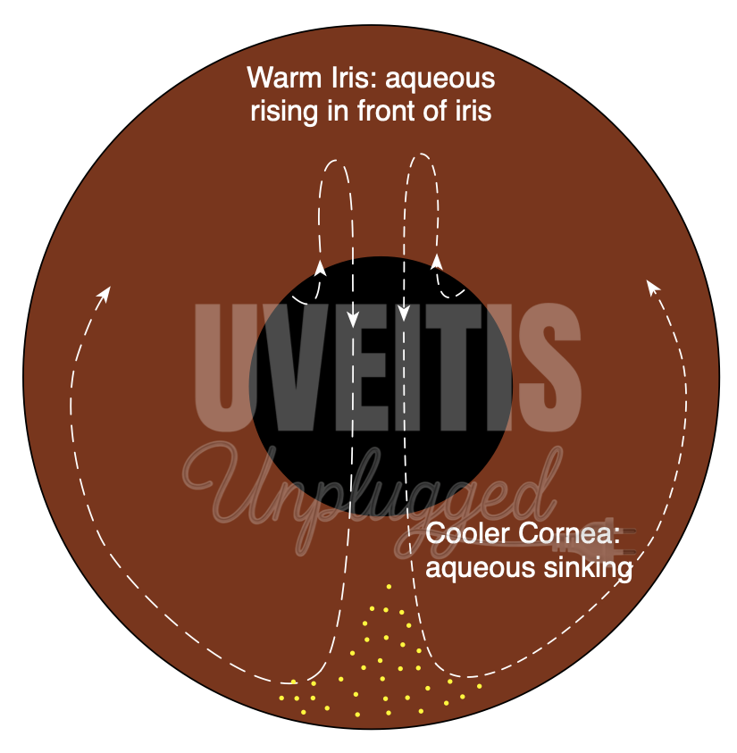
Keratic precipitates: Keratic precipitates (KPs) are cellular deposition on corneal endothelium during uveitis.
Pathogenesis of KP: Migration of inflammatory Cells across the iris and ciliary vessels to aqueous humour >> In AC, these cells come across a convection current (due to temperature difference between inflamed warm iris & relatively cooler cornea) >> They start moving in AC because of this convection current >> Because of the altered nutrition (inflammatory products in aqueous), corneal endothelium becomes sticky and may desquamate in places. And circulatory cells due to centrifugal convection current (convection currents in the anterior chamber that rise along the warm iris and fall along the cool cornea) and gravity often sticks on the corneal endothelium, which are termed as Keratic precipitates (KP).
|
Distribution of KPs |
|
|
Arlt's triangle |
Base down triangular area in the lower part of the cornea |
|
Turk’s Line |
Arranged in a linear fashion (vertical line) |
|
Scattered over whole cornea |
Viral infections, and Fuchs' heterochromic iridocyclitis |
|
Type of cells |
|
|
Acute inflammation |
Polymorphonuclear leucocytes |
|
Chronic inflammation |
Lymphocytes Plasma cells and pigment cells |
|
Morphology of KPs |
|
|
Fresh KP |
White and round |
|
Old KP |
Pigmented, faded, irregular crenated margins |
|
Types of KPs |
|
|
Mutton fat KPs |
Large KPs, clusters of macrophages and epithelioid cells, pathognomonic of granulomatous inflammation. |
|
Fine KPs |
Viral infections, Nongranulomatous uveitis |
|
Stellate KPs |
Fuchs' heterochromic iridocyclitis |
Band-shaped keratopathy: Deposition of calcium hydroxyapatite in Bowman's layer can be seen in conditions like chronic uveitis juvenile idiopathic arthritis. It typically begins at the periphery of the interpalpebral region and spread centrally. If the visual axis is involved, it can cause a marked diminution of vision. On slit-lamp examination, small clear dot-like areas are observed, giving a "Swiss cheese" appearance to band keratopathy. These clear, small dot-like areas represent the location where corneal nerves penetrate Bowman's layer.
Scar or Ulceration:
- Cornea should be examined properly for superficial or deep scar, dendritic epithelial keratitis to detect keratouveitis.
- Stromal thinning and epithelial ulceration due to inflammation may be observed in peripheral ulcerative keratitis (PUK). PUK may also be the first sign of systemic necrotizing vasculitis like rheumatoid arthritis, Wegener’s Granulomatosis, etc.
D. Examination of pupil:
- Pupillary reaction in acute cases of iritis or iridocyclitis becomes sluggish or abolished and constriction of pupil occurs.
- Often the exudates released during acute inflammation causes plastering of the posterior surface of iris with anterior capsule of the lens. These iridolenticular adhesions are known as posterior synechiae.
- Festooned pupil: Due to the application of cycloplegic topicals, often some parts of the posterior synechiae dilates thus making the pupil festooned shaped.
- In cases of severe anterior chamber inflammation, the pupillary margin may become plastered to the lens capsule called annular or ring synechiae or seclusio pupillae.
- And if the pupillary aperture is blocked by the exudates forming a membrane over it then it is called occlusio pupillae.
- Similarly adhesion between iris and the cornea near the anterior chamber angle can occur due to inflammatory process and known as peripheral anterior synechiae (PAS). Posterior synechiae can lead to pupillary block glaucoma, and peripheral anterior synechiae can cause secondary angle-closure glaucoma
- Afferent pupillary defect is seen in patients with Neuroretinitis.
E. Examination of Iris:
- In acute iritis or iridocyclitis, the iris loses its normal pattern due to imbibition of inflammatory exudates in it and such iris is often called muddy iris.
- Accumulations of inflammatory cells in the iris or on iris surface can be clinically noted as iris nodule. Nodules seen in pupillary borders are known as Koeppe nodule and nodules on the iris surface is known as Busacca's nodule. Iris nodules are seen in granulomatous uveitis. Various types of iris nodules are as follows:
|
Name |
Location |
Condition |
|
Koeppe nodules |
Pupillary border |
Granulomatous Uveitis |
|
Busacca nodules |
Anterior surface of the iris |
Granulomatous Uveitis |
|
Berlins nodules |
Angle of anterior chamber |
Sarcoidosis |
|
Lisch nodules |
Surface of the iris |
Neurofibromatosis type-I, not related to uveitis |
- Following are the differentiating features between Koeppe nodules and Busacca nodules
|
Koeppe’s nodule |
Bussaca’s nodule |
|
|
- Also, one must be able to differentiate between Iris nodules from iris Granuloma
|
Iris nodules |
Iris granulomas |
|
Iris nodules are accumulated deposits of epithelioid cells and lymphocytes that have been deposited onto the iris without tissue destruction |
Granulomas are whitish grey lesions and are made up of mononuclear phagocytes, epithelioid cells and giant cells located within iris stroma & heal by fibrosis |
- Comparison of the colour of iris between two eyes can detect heterochromia of iris which can be either hypochromic (abnormal eye is lighter than fellow eye) as seen in Fuch’s heterochromic c iridocyclitis or hyperchromic (abnormal eye is darker than fellow eye) as seen in melanosis of iris.
- Iris atrophy is a characteristic feature of herpetic uveitis. Herpesviruses generally produces sector iris atrophy due to an occlusive vasculitis
Causes of iris atrophy
- Viral infections
- Anterior segment ischemia,
- Leprosy,
- syphilis
- Previous attacks of angle-closure glaucoma.
- Iatrogenic (previous intraocular surgery)
- In syphilis, dilated, hyperemia of the iris vessels are noted, which is known as roseolae
F. Intraocular pressure (IOP):
- Patients with acute iridocyclitis have low IOP due to infiltration of the ciliary body by inflammatory cells, leading to aqueous production reduction. Release of prostaglandin during inflammation can also cause a reduction in IOP.
- Sometimes, patients with uveitis can present with a significant rise in IOP. Conditions like herpes simplex uveitis, herpes zoster uveitis, PosnerSchlossman syndrome, and toxoplasmosis can present with rise of IOP in acute cases.
The cause of rise of intraocular pressure in uveitis can be due to:
- clogging of trabecular meshwork with inflammatory cells,
- inflammation of trabecular meshwork fibres (“trabeculitis”),
- Peripheral anterior synechiae
- Pupillary block from posterior synechiae and
- Corticosteroid-induced IOP rise (steroid responder).
- In case of long-standing uveitis, extensive membrane formation over ciliary body or ciliary body detachment can cause ocular hypotony and eventual phthisis bulbi.
G. Examination of lens:
- Posterior subcapsular cataract is a common complication after long-standing uveitis as well as chronic corticosteroid therapy.
H. Examination of vitreous:
- Cells in the anterior vitreous or retrolental space should be looked for after pupillary dilation. Though there is no standard grading system for vitreous cells, documentation of this finding is important for the follow-up of a uveitic case.
- Vitreous should be carefully examined with a 78/90D and also with indirect ophthalmoscope with an indentation for snowball opacities, snow-banking in pars plana region and vitreous strands
- For classifying vitreous haze, SUN classification has adopted The National Eye Institute system for grading vitreous haze with the proviso that the designation “trace” be recorded as 0.5+.
|
0 |
nil |
|
0.5+ |
trace |
|
1+ |
Few opacities, mild blurring |
|
2+ |
Significant blurring, but still visible |
|
3+ |
Optic nerve visible, no vessels visible |
|
4+ |
Dense opacity obscures optic nerve head |
Table National eye institute grading system for vitreous haze (adopted by SUN classification)
I. Examination of Fundus:
- Optic disc should be carefully examined with the help of slit lamp biomicroscopy. Disc hyperemia, disc oedema, or optic neuritis is seen in various uveitic conditions. Glaucomatous damage to the optic disc due to Secondary glaucoma, Neovascularization of the optic disc, optic disc granuloma and optic atrophy may also occur in uveitic patients.
- Examination of retinal vasculature for vasculitis, vascular sheathing, and accumulation of inflammatory cells around vessels is important. Vascular sheathing is seen as white parallel lines along vessels. Sometimes inflammatory exudates are seen around the vessels in patients with sarcoidosis, known as candle-wax drippings. Also it is important to determine whether retinal veins, retinal arteries, or both are affected as it can help in differential diagnosis of the uveitic entity.
|
Arterial involvement (arteritis) |
Venous involvement (phlebitis) |
|
|
- Careful examination of the posterior segment can reveal inflammatory patches in the fundus. It is important to distinguish such lesions whether it involves retina or choroid or both. Sometimes these lesions are associated with subretinal fluid or localized haze in vitreous.
|
Retinitis |
Choroiditis |
|
|
- Often a patient can present with a retinal detachment. Exudative retinal detachments can be seen in a number of ocular inflammatory diseases like Vogt–Koyanagi–Harada syndrome. One should be able to distinguish rhegmatogenous retinal detachment from such cases, which requires surgical management. Sequelae to vasculitis can lead to the development of traction retinal detachment and should be dealt properly.
- Meticulous examination of the fovea with slit lamp biomicroscopy often helps to identify cystoid macular edema, choroidal neovascular membrane or sight-threatening inflammatory lesions like serpiginous choroiditis. Cystoid macular edema is a common in patients with uveitis which if long standing can lead to formation of macular hole.