Anatomy of Uveal Tract: Iris
Our eyeball consists of three coats. The uvea is the middle vascular coat of the eyeball. The name 'uvea' originates from the Latin word for 'grape’. Why is it called a grape? Because if the stem is removed from a grape, the resulting hole resembles the pupil, with the grape mimicking the shape of an eyeball. From anterior to posterior, the uvea or uveal tract can be divided into three parts: the iris, the ciliary body, and the choroid.
The uveal tract is a vascular layer rich in pigments and composed of various mesenchymal cell types. These include fibroblasts, melanocytes, and cells that support the vascular structure, originating primarily from the neural crest (mesoectoderm) and, to a lesser extent, from the mesoderm, which includes vascular endothelial cells.
Iris:
The iris is the most anterior part of the uveal tract. It is a thin, circular structure that forms a diaphragm in front of the crystalline lens. The word 'iris' originates from a Greek word. In Greek mythology, 'Iris' is the name of the goddess of the rainbow. The diaphragm formed by the iris contains a central aperture known as the pupil. The pupil's location is not exactly central; it is slightly nasal to the centre. The pupil controls the amount of light that enters the eye. The normal size of the pupil is 3-4 mm.
The anterior surface of the iris can be divided into a pupillary zone and a ciliary zone by an imaginary circular ridge called the collarette, located 1.5 mm away from the pupillary margin.1,2 The iris is attached to the middle of the anterior surface of the ciliary body and divides the space in front of the lens into the anterior and posterior chambers. The average diameter of the iris is 10-11 mm. It is thickest at the collarette, located approximately 1.5 mm from the pupillary margin, and thinnest at the iris root, the part of the iris that joins with the ciliary body. The thickness of the iris root is approximately 0.5 mm. During blunt trauma, the iris root is most vulnerable and can tear from its attachment, leading to iridodialysis.
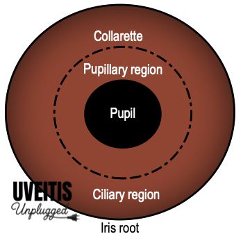
The collarette, an imaginary zone, holds several anatomical importance. First, the collarette is the site of foetal pupillary membrane attachment. Persistence of a pupillary membrane occurs when the anterior portion of the tunica vasculosa lentis fails to be absorbed during foetal development. Second, in the collarette, the iris sphincter muscle and the dilator muscle overlap. Third, the collarette is the region where anastomoses between the arterial and venous arcades occur, forming the minor vascular circle of the iris.
Pupillary Zone:
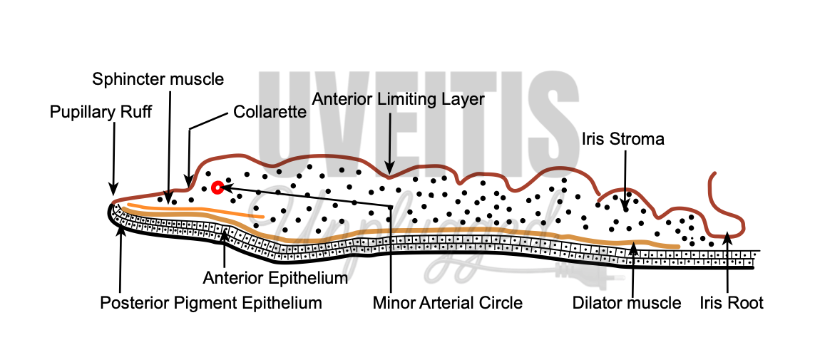
The pupillary zone extends from the pupillary margin to the collarette. The pupillary zone is relatively flat. The pupillary margin is marked by a dark border, known as the pupillary ruff. The pupillary ruff is the anterior termination of the pigmented layer that lines the posterior surface of the iris. Under normal conditions, when the pupil is not dilated, its margin rests on the lens. In the absence of this support, as in aphakic patients, the pupil becomes tremulous
Ciliary Zone:
The ciliary zone of the iris extends from the collarette to the iris root. In this area, there are depressions or pits arranged in rows, known as crypts. Crypts are found in two locations: near the collarette and at the periphery of the iris. Those near the collarette, known as Fuchs’s crypts, are relatively larger, while a few are seen at the periphery of the iris. In laser iridotomy, openings are created in the areas of iris crypts because these are the thinnest areas, requiring less energy.
Posterior surface of iris:
The posterior surface of the iris presents a more uniform appearance compared to the anterior surface, characterized by its darker coloration and numerous radial contraction folds. Additionally, circular folds are visible on this surface.
Microscopic structure:
1. Anterior limiting layer
The anterior surface of the iris is not covered by an epithelium but by an incomplete layer known as the anterior limiting layer. As a result, the aqueous humour freely permeates the iris stroma. The anterior limiting layer represents the most anterior condensation of the iris stroma and consists of fibroblasts and melanocytes. These cells are arranged in a meshwork: fibroblasts are located on the surface, and melanocytes are beneath them.
The colour of the iris is determined by how much melanin, a type of pigment, is in the melanocytes. Melanocytes are found in anterior limiting membrane and superficial stroma of the iris. These melanocytes seem to acquire their melanin content in early childhood, which is genetically determined. However, some evidence suggests that the melanin content of some melanocytes is subject to adrenergic regulation even after childhood. (3)
The iris surface looks smooth in dark-coloured eyes because heavily pigmented stroma. In contrast, the surface of blue-coloured eyes appears more irregular due to less pigmented stroma. To simplify, if there is less pigment in the iris, it appears blue or green because it absorbs longer wavelengths of light and reflects shorter ones.(3) Certain conditions can lead to decreased iris pigmentation, such as Horner’s syndrome and Fuchs’ Uveitis. On the other hand, the use of the prostaglandin analogue latanoprost can lead to an increase in iris pigmentation. (3)
2. Iris Stroma:
Iris stroma forms the main bulk of iris tissue and contains sphincter pupillae, dilator pupillae muscles, vessels and nerves.
Cells in Iris Stroma
|
Pigmented cells |
Non-pigmented cells |
|
Melanocytes + Clump cells |
Fibroblasts + Lymphocytes + Macrophages + Mast cells |
Fibroblasts are the most prominent cells in iris stroma. These cells are located in close association with blood vessels, muscles, and nerves. Melanocytes can be found around the adventitia of blood vessels. Melanocytes contain mitochondria, smooth and rough endoplasmic reticula, free ribosomes and melanin granules in various stages of development. (5) They have long processes with the help of which they form plexuses with fibroblasts and adjacent melanocytes. Clump cells (also called clump cells of Koganei) are large, round, pigmented cells, located in the pupillary portion of the stroma, mainly near the sphincter muscle. (6) There are two types of clump cells: (5)
- Type I cells are numerous and believed to be “altered macrophages” and they act as scavengers of free pigments, present within the iris.
- Type II cells are less in number and are thought to represent smooth muscle cells in arrested stages of development.
Muscles in Iris Stroma
|
Sphincter pupillae |
Dilator pupillae |
|
Located in Pupillary Zone of posterior stroma encircling pupil |
Located in Ciliary Zone between Stroma and Posterior Pigment Epithelium, extending from Iris Root to Sphincter muscle |
|
Nerve Supply: Parasympathetic, via short ciliary nerves |
Nerve Supply: Sympathetic, via long ciliary nerves |
The sphincter pupillae muscle is a circular muscle, 0.75 to 1 mm wide, composed of smooth muscle cells. The muscle is 0.1 to 1.7 mm in thickness and is considerably thicker than the dilator papillae. It encircles the pupil and is located in the pupillary zone of the stroma. The sphincter muscle is firmly adherent to the surrounding stroma of iris. The sphincter pupillae retains its function even if severed radially (after sphincterotomy) because of its unique distribution of fibres.1 Sphincter muscle is composed of spindle-shaped cells that are oriented parallel to the pupillary margin, so, contraction of the sphincter causes the pupil to constrict (a process known as miosis). The muscle is innervated by the parasympathetic system. (7)
The dilator pupillae muscle extends from the iris root to a point in the stroma below the midpoint of the sphincter. A dense band of connective tissue separates the sphincter and dilator muscles from each other. However, near the termination of the dilator muscle, small projections insert into the sphincter. (1) Because of the radial arrangement of the fibres of the muscle, contraction of the dilator pupillae muscle pulls the pupillary portion toward the root, thereby enlarging the size of pupil (a process known as mydriasis). The dilator pupillae muscle is sympathetically innervated. (7)
Blood vessels in iris stroma:
Iris vessels include arterioles, venules, and capillaries. The iris arteries are branches of major circle of the iris, located in the ciliary body near the iris root. The iris vessels usually follow a radial course from the iris root to the pupil margin. These vessels are surrounded by a dense network of collagenous fibrils which is embedded into the collagen network of the stroma. Such arrangement of collagen network prevents the iris vessels from kinking and compression during the extensive iris movement during constriction and dilatation of pupil. Iris capillaries have relatively large diameter, and their endothelium is nonfenestrated and is surrounded by a basement membrane, associated pericytes, and a zone of collagenous filaments. (8) The intima of these capillaries has no internal elastic lamina. Iris veins have very thin walls consisting of endothelium surrounded by a thin layer of collagen. Capillaries are formed by a single layer of unfenestrated epithelium, which forms a part of the blood-aqueous barrier. (9)
Nerves in iris stroma:
Iris nerves are usually unmyelinated; however some nerves are found be enclosed by Schwann cells. (7)
3. Iris Pigment Epithelium:
Posterior surface of Iris has two mono layers of epithelium that lie beneath the stroma – both are pigmented, and epithelial apical portion of anterior pigment epithelium is in close apposition with the apical surface of the posterior pigment epithelium. Such apex-to-apex arrangement is to be seen between the pigmented and nonpigmented epithelium of ciliary body. They form and maintain the blood-aqueous barrier.9 The posterior cells of the iris pigment epithelium are heavily pigmented and prevent light from reaching the retina.10,11 Conversely, the anterior iris pigment epithelial cells undergo modification to constitute the dilator muscle.
The cells in anterior pigment epithelial layer of the iris have two distinct portions.
- Muscular basal portion anteriorly (lies next to stroma): composed of elongated, contractile, smooth muscle fibres. These muscle fibers extend into the stroma, forming three to five layers of dilator muscle fibers joined by tight junctions.
- Epithelial apical portion posteriorly (lies above posterior pigment epithelium of iris): composed of pigmented cuboidal epithelium where cells are joined by tight junctions and desmosomes.
The anterior iris epithelium continues posteriorly as the pigmented epithelium of the ciliary body.
Posterior pigment epithelial cells are rectangular or pyramidal in shape with round cell nucleus and their cytoplasm contains large pigment granules. The cells of the posterior pigment epithelium are more heavily pigmented than anterior pigment epithelium. The cells are joined to each other by maculae adherens and occludens. These cells rest on a thin layer of basement membrane situated posteriorly.
References:
- Bloom J, Motlagh M, Czyz CN. Anatomy, Head and Neck: Eye Iris Sphincter Muscle. In: StatPearls. StatPearls Publishing; 2024. Accessed April 11, 2024. http://www.ncbi.nlm.nih.gov/books/NBK532252/
- Li S, Liang L. Protruding Iris Collarette. N Engl J Med. 2017;376(11):1064. doi:10.1056/NEJMicm1516330
- Imesch PD, Wallow IH, Albert DM. The color of the human eye: a review of morphologic correlates and of some conditions that affect iridial pigmentation. Surv Ophthalmol. 1997;41 Suppl 2:S117-123. doi:10.1016/s0039-6257(97)80018-5
- Gartner S, Henkind P. Neovascularization of the iris (rubeosis iridis). Surv Ophthalmol. 1978;22(5):291-312. doi:10.1016/0039-6257(78)90175-3
- McMenamin PG. The distribution of immune cells in the uveal tract of the normal eye. Eye (Lond). 1997;11 ( Pt 2):183-193. doi:10.1038/eye.1997.49
- Wobmann PR, Fine BS. The clump cells of Koganei. A light and electron microscopic study. Am J Ophthalmol. 1972;73(1):90-101. doi:10.1016/0002-9394(72)90311-x
- McDougal DH, Gamlin PD. Autonomic control of the eye. Compr Physiol. 2015;5(1):439-473. doi:10.1002/cphy.c140014
- Bill A. Blood circulation and fluid dynamics in the eye. Physiol Rev. 1975;55(3):383-417. doi:10.1152/physrev.1975.55.3.383
- Freddo TF. A contemporary concept of the blood-aqueous barrier. Prog Retin Eye Res. 2013;32:181-195. doi:10.1016/j.preteyeres.2012.10.004
- Wang X, Xiong K, Lu L, et al. Developmental origin of the posterior pigmented epithelium of iris. Cell Biochem Biophys. 2015;71(2):1067-1076. doi:10.1007/s12013-014-0310-0
- Thumann G. Development and cellular functions of the iris pigment epithelium. Surv Ophthalmol. 2001;45(4):345-354. doi:10.1016/s0039-6257(00)00195-8