From Bloodletting to Breakthroughs: How We Fought Uveitis
A peek into the history of how we treated uveitis, originally published in AIOS Times.
Ancient Mysteries
Imagine you are suffering from red, painful eyes and you decide to consult a well-known doctor in your neighbourhood. As you walk into his chamber, he merely looks at your eyes. Seeing the fiery redness and a white streak in the lower part of your eye—what we now know as a hypopyon, the doctor says, “It’s uveitis.” He quickly applies a lotion and asks you to lie down. You notice him moving toward a large water tank at the side of the room. He carefully reaches in and pulls out two large leeches. Two assistants hold your arms firmly as the doctor gently places the leeches near your eyes—one on each side. Torniquets are tied around your arms and legs, and small cuts are made to let blood flow freely. Strange as it may sound today, this was common medical practice in the 19th century. In fact, back in 1806, Scarpa, a well-known physician of his time, described in detail how he treated hypopyon uveitis in this very way. (1) Bloodletting—by knife or by leech—was believed to draw out illness. It was the standard treatment for nearly every disease.
From the ancient Egyptian papyri to the writings of Hippocrates, uveitis has continued to puzzle doctors — just as it still does today. Many forms of uveitis were observed and described by early physicians, even if they didn’t fully understand what they were seeing. For example, Hippocrates wrote about symptoms of what we now know as Behçet’s Syndrome in his Third Book of Endemic Diseases. But it wasn’t until the 1930s that modern medicine recognized it clearly, thanks to the work of Benediktos Adamantiades and Hulusi Behçet. Although early knowledge of eye anatomy was often limited or incorrect, we find descriptions of the uveal tract in the work of Andreas Vesalius, the father of anatomy. Even earlier, the Roman physician Galen used the terms ‘choroid’ and ‘choroiditis’. Medicine, for centuries, thrived on the art of observation. Long before modern diagnostics and therapeutics, ancient physicians relied on the art of observation to identify diseases, even without understanding their pathophysiology—testament to the sharpness of their clinical eye. For example, in 1567, Juan de Vigo recognized the eye symptoms of syphilis and accurately referred to it as the ‘great imitator.’
One thing remained true across all eras: while doctors could recognize the disease, they didn’t know what caused it. So treatment was mainly aimed at easing symptoms, not curing the root problem. As time passed, the unknowns became fewer, but uveitis has always remained a mysterious and complex condition — from ancient times to the modern age.
The Age of Steroids
Deborah I. Friedman, a professor of neurology and ophthalmology at the University of Texas, once said, “No one is allowed to die or go blind without a trial of steroids!”—a powerful reminder of how crucial corticosteroids are in medical treatment today. During the COVID pandemic, when the world was reeling under the tsunami of cytokine storm, corticosteroids rose as a beacon of hope, saving countless lives. The discovery of corticosteroids truly felt like a miracle for managing inflammation also. Before corticosteroid was discovered, treating inflammation was not easy.
In the early twentieth century, doctors used strange and risky methods like injecting milk into muscles or giving triple typhoid H antigen through the vein to raise the body’s temperature to around 40°C, which could stimulate natural cortisol release—but this method was unpredictable and sometimes deadly. (2) We can take the example of rheumatoid arthritis, one of the most important and common systemic rheumatic diseases, which caused severe pain, deformities, and disability—especially in young people—before the discovery of corticosteroids. Ao naturally, scientists were desperate to find a cure for this disease. One such scientist was Philip Showalter Hench from the Mayo Clinic, who was very observant and believed that the adrenal glands had something important to offer in treating this disease. He initially tried using lactophenin, a medicine known to induce jaundice, based on reports that jaundice seemed to improve rheumatoid arthritis. He also noticed that women with rheumatoid arthritis often felt better during pregnancy—something we now know is due to higher cortisol levels during weeks 30 to 32. Since the 1880s, following the work of Addison and Brown-Séquard, it had been well established by then that animals without adrenal glands would rapidly die when subjected to even mild stress. Combining these observations, Hench hypothesized that some endogenous substance — likely produced during pregnancy or stress — was reducing inflammation. At the same time, Edward C. Kendall, a chemist working at the animal research facility of same clinic, was studying adrenal glands from cows. After processing many kilograms of bovine adrenal tissue, they finally extracted just 5 mg of a substance they called Compound E. Hench selected a 29-year-old woman who had been suffering from severe rheumatoid arthritis for four years and was bedridden. Compound E (50 mg) was injected twice a day for 5 days in September 1948. Amazingly, just two days after the injection, she started feeling better, and by the third day, she was able to get out of bed—something she hadn’t done in months. This compound E was cortisone and the rest is history. Two years later, in 1950, Hench and his colleagues received the Nobel Prize—by then, corticosteroids had already entered the field of ophthalmology. Gordon and McLean had published a paper in 1950, demonstrating how effective corticosteroids were in the treatment of eye inflammation.3 That same year, Kendall and his colleagues discovered a slightly different version of Compound E, which they named Compound F. This new version was more soluble and required smaller doses. This compound was none other than hydrocortisone, cousin of cortisone! Hench began using it as a local joint injection to reduce the side effects of cortisone. The idea of injecting steroids directly into the eye came later, and quite unexpectedly. In 1977, a dermatologist was treating a 35-year-old man with a steroid injection in his upper eyelid while using a special device called a Dermojet.4 By accident, the needle pierced the eye and delivered 0.1 ml of triamcinolone acetonide (40 mg/ml) inside the vitreous cavity. Surprisingly, the eye didn’t suffer any serious damage. This encouraged further tests, first in rabbit eyes, and soon led to a new way of treating eye diseases through intravitreal steroid injections—an approach now widely used now a days.
The War that Gifted Medicine
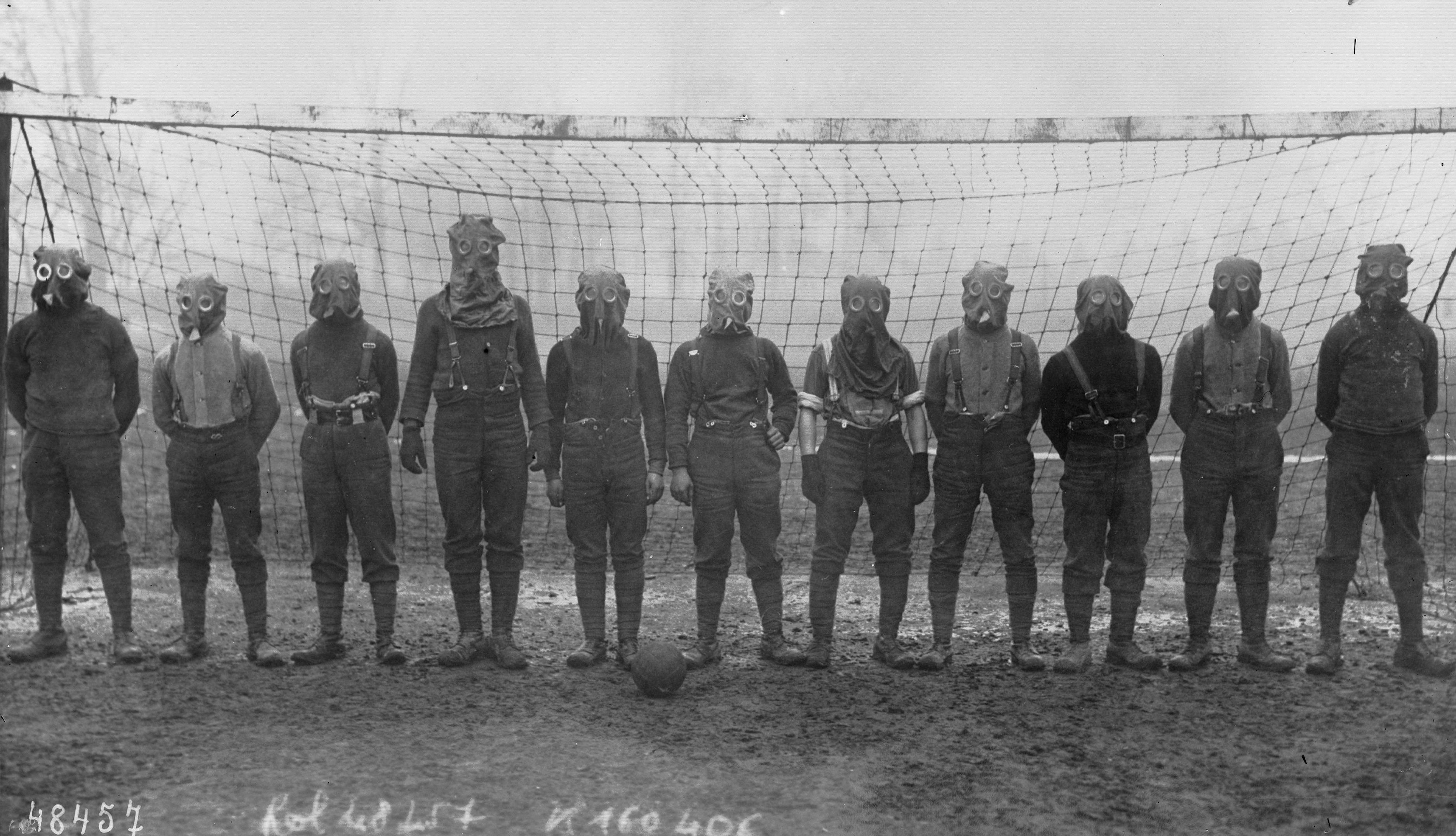
Every storm, no matter how destructive, often leaves behind a seed of renewal in the form of unexpected gifts. If we look at World War II, despite its devastation, it also sparked ideas that led to some of the greatest advancements in medicine. One such example comes from the experience of treating Royal Air Force pilots who had eye injuries caused by fragments of their aircraft canopies made of acrylic, or PMMA. While caring for these soldiers, Sir Harold Ridley realized the potential of PMMA, this inert material and later went on to invent and implant the first intraocular lens—a groundbreaking moment in ophthalmology. Another lesser-known but equally important outcome of World War II was the development of drugs to fight cancer and the beginning of immunosuppressive therapy. Mustard gas, a derivative of nitrogen or sulphur mustard, was first developed in the 19th century and used as a chemical weapon during World War I and much widely during World War II. Scientists observed that exposure to mustard gas caused suppression of bone marrow and a decrease in white blood cells, leading to conditions like leukopenia and destruction of lymphoid tissue in affected individuals. These harmful effects sparked the idea that such chemicals could be used to suppress abnormal cell growth. In 1953, researchers found that aromatizing mustard gas could reduce its toxicity. This led to the development of chlorambucil. In 1958, another major step was taken when scientists added phenylalanine to the molecule, creating cyclophosphamide, which improved its uptake by human cells. These discoveries turned a weapon of mass destruction into a powerful tool to treat cancer. Later, this drug found its place in managing immune-related conditions as well. But you have to agree—doctors who treat uveitis are really smart ! Much before the invention of cyclophosphamide and chlorambucil, Roda Perez published an article on the treatment of uveitis in 1952. The topic? Nitrogen mustard therapy for uveitis of unknown etiology. (5)
References:
- Scarpa A. Practical Observations on the Principal Diseases of the Eyes. London: Strand; 1806. pp. 292-321.
- Benedek TG. History of the development of corticosteroid therapy. Clin Exp Rheumatol. 2011 Sep-Oct;29(5 Suppl 68):S-5-12. Epub 2011 Oct 21. PMID: 22018177.
- Gordon DM, McLean JM. Effects of pituitary adrenocorticotropin hormone (ACTH) therapy in ophthalmologic conditions. JAMA. 1950; 142:1271-6.
- Perry HT, Cohn BT, NAuheim JS. Accidental intraocular injection with Dermojet syringe. Arch Dermatol. 1977 Aug;113(8):1131. PMID: 889347.
- Roda Perez E. El tratamiento de las uveitis de etiologia ignota con mostaza nitrogenada [Nitrogen mustard therapy of uveitis of unknown etiology]. Rev Clin Esp. 1952 Feb 15;44(3):173-80. Undetermined Language. PMID: 14949545.