Anatomy of Uveal Tract: Ciliary body
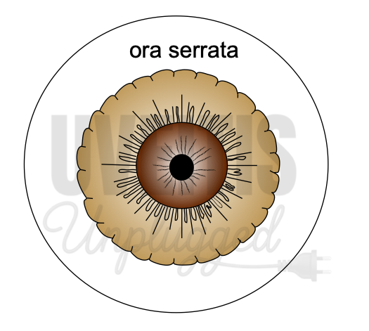
The ciliary body is the middle part of the uveal tract. It is a ring-shaped (slightly eccentric) structure that projects posteriorly from the scleral spur, with a meridional width varying from 5.5 to 6.5 mm. (1),(2) It is brown in colour due to melanin pigment. Anteriorly, it is confluent with the periphery of the iris (iris root), and the anterior part of the ciliary body forms a part of the anterior chamber angle. Posteriorly, the ciliary body has a crenated or scalloped periphery, known as the ora serrata, where it is continuous with the choroid and retina. The ora serrata exhibits forward extensions, known as dentate processes, which are well-defined on the nasal side and less so temporally. (1),(2) The ciliary body has a width of approximately 5.9 mm on the nasal side and 6.7 mm on the temporal side.
Extension of the ciliary body
On the outside of the eyeball, the ciliary body extends from a point about 1.5 mm posterior to the corneal limbus to a point 6.5 to 7.5 mm posterior to this point on the temporal side and 6.5 mm posterior on the nasal side.
Parts of Ciliary body:

Ciliary body, in cross-section, is a triangular structure (in diagram # it can be compared as ∆ AOI). The outer side of the triangle (O) is attached to the sclera with suprachoroidal space in between. The anterior side of the triangle (A) forms part of the anterior & posterior chamber. In its middle, the iris is attached. The inner side of the triangle (I) is divided into two parts. The anterior part (2 mm) is known as pars plicata (corona ciliaris) and the posterior smooth (4 mm) is known as pars plana (orbicularis ciliaris). (1),(2)
Pars plicata:
The pars plicata is the portion of the ciliary body that contains the ciliary processes. Ciliary processes are finger-like projections, which extend into the posterior chamber. The regions between ciliary processes are called valleys of Kuhnt. Ciliary processes are approximately 70 to 80 in number. A ciliary process measures approximately 2 mm in length, 0.5 mm in width, and 1 mm in height.
Pars plana:
As discussed earlier, pars plana is the flat or smooth part of the ciliary body. It terminates at the ora serrata, which is the transitional zone between the ciliary body and choroid. Histologically, the pars plana consist of a double layer of epithelial cells: the inner, nonpigmented epithelium, which is continuous with neurosensory retina; and the outer, pigmented epithelium, which is continuous with the retinal pigment epithelium (RPE). (1),(2),(3)
The pars plana is a relatively avascular zone, which is important surgically in the pars plana approach to the vitreous space. Pars plana provides surgical access to the vitreous and retina.
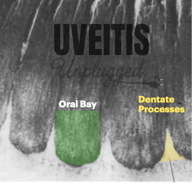
Ora serrata: the transition zone
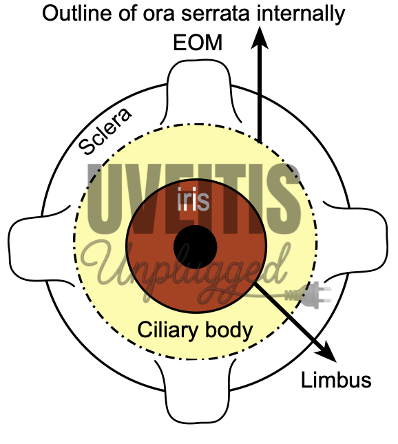
The ora serrata can be termed as the anterior border of the neurosensory retina. Ora serrata shows forward extensions into the retina, which are well-defined in the nasal side and less so temporarily. Dentate processes are “teeth-like” extensions of neurosensory retina into pars plana. There are approximately 20 to 30 dentate processes per eye. Ora bays are rounded extensions of the pars plana. (1),(2) Topographically, ora serrata corresponds to the insertion of the medial and lateral rectus muscles.
Lens and ciliary body:
The zonules course from ciliary body to the lens. Some of these zonular fibers insert into the internal limiting membrane of the pars plana region and travel forward through the valleys (valleys of Kuhnt) between the ciliary processes. It can be mentioned here that the distance between equator of lens and ciliary body is approximately 0.5 mm.
Layers of ciliary body:
From inside to outside (from sclerad to vitread), ciliary body consists of the following four layers.
1. Ciliary epithelium:
Ciliary epithelium consists of two layers that cover the inner surface of the ciliary body (don’t confuse !! from vitreous side, it is external side). These two layers are representative of two layers of optic cup embryologically.
1A. Non pigmented epithelium (NPE) of ciliary body:
NPE of ciliary body extends from iris root to ora serrata. It begins as a continuation of posterior pigmented epithelium of iris near iris root. In nonpigmented epithelium, cells become smaller and there is significant decrease in melanin granules in the cells. At pars plicata, these cells are cuboidal, which gradually becomes columnar with increasing age. At ora serrata, the NPE continues as sensory retina.
The internal limiting membrane is secreted by the basal lamina of the non-pigmented epithelium on its basal surface, which is on the vitreal side. It is the continuation of inner basement membrane of the iris and continues posteriorly as inner basement membrane of retina. It gives origin to parts of the suspensory lens ligament.
1 B. Pigmented epithelium of ciliary body:
The cells of the pigmented epithelium are 8 to 10 micronm wide and contain large pigment granules. These pigment granules are three to four times larger than those of the choroid and retina. These cells are rich in organelles and are very active metabolically. The cells of pigment epithelium secrete basement membrane which continues posteriorly with the retinal pigment epithelium (RPE).
Ciliary epithelium: metabolic activity
The cells of both the ciliary epithelium have a greater number of mitochondria and thus they have a higher degree of metabolic activity, with a significant role in the active secretion of aqueous humour. As the name suggests, pigmented epithelium cells have large melanosomes which occupy almost whole of the cytoplasm. It has to be kept in mind that because of the unique apex-to-apex configuration of the cells of the non-pigmented and pigment epithelium of ciliary body, basement membrane of the non-pigmented epithelium faces the posterior chamber whereas basement membrane of the pigmented epithelium of ciliary body rests on the stroma of the ciliary body .
The cellular junctions found between the pigmented and non-pigmented epithelia are zonulae occludentae, gap junctions, desmosomes, and puncta adherentia. These connecting structures are important for the secretory role of the ciliary processes.
2. Ciliary stroma:
Ciliary stroma consists of bundles of loose connective tissue. Ciliary stroma contains blood vessels, nerves and ciliary muscle. Ciliary stroma continues anteriorly with iris stroma and continues posteriorly with choroidal stroma after thinning out at pars plana.
Blood vessels in ciliary stroma: Major arterial circle of iris
Major arterial circle of iris, formed by the anastomosis of long posterior ciliary arteries and anterior ciliary arteries, is located in ciliary stroma near the iris root just in front of the circular portion of ciliary muscle. Ciliary stroma also consists of numerous capillaries which are fenestrated and large in size. The capillaries are more in number in ciliary processes, making them the most vascular organ of the eye.
Muscle in ciliary stroma: ciliary muscle.
Ciliary muscle is a nonstriated or smooth muscle primarily situated in the anterior two thirds of the ciliary body stroma.4 The muscle has three parts
- Outer longitudinal or meridional portion (Brücke's muscle): This is the most external part (nearest to the sclera) of the ciliary muscle. This part of the muscle is v-shaped, the base of the v is attached to the scleral spur and limbs are inserted into the stroma of choroid.
- Middle oblique portion (also called reticular or radial): This part of the muscle also originates from the scleral spur and the muscle fibres are attached to the collagenous substances near ciliary processes.
- Inner circular portion (Müller's muscle): Here the muscle bundles are circular in shape (that’s why it is also called annular part of ciliary muscle) and act as a sphincter. It lies close to the periphery of lens and is embedded in ciliary stroma near the major arterial circle of iris.
Contraction of the ciliary muscle, especially of the longitudinal and circular fibers, pulls the ciliary body forward during accommodation. This forward movement of ciliary body relieves the tension in the suspensory lens ligament (zonules), making the elastic lens more convex and thereby helping the eye in accommodation by increasing the refractive power of the lens.3,4
Ciliary muscle is innervated by the autonomic nervous system by parasympathetic postganglionic fibres derived from the oculomotor nerve. The nerve fibres reach the muscle via the short ciliary nerve. The parasympathetic stimulation activates the muscle for contraction, whereas sympathetic innervation likely has an inhibitory effect.
Parasympathetic fibers, coming from the Edinger Westphal nucleus with the oculomotor nerve, are mixed with nerve fibres from the ciliary ganglion and form a plexus in the ciliary muscle.
Kindly note that while classifying the different parts of ciliary muscle, the term outer is used as external, meaning nearer to sclera or the outer side.
Sympathetic fibers from the cervical sympathetic trunk, synapse in the superior cervical ganglion, and run to the ciliary muscle via the long ciliary nerve. The sensory fibres, coming from the nasociliary branch of the trigeminal nerve, also run from the long ciliary nerve to the ciliary body and terminate in the ciliary muscle.
Supraciliary lamina:
Supraciliary lamina is the outermost layer of ciliary body which lies adjacent to the sclera. It is composed of loose connective tissue with collagen strands, fibroblasts and melanocytes. Some of the collagen strands merge with scleral collagen. Because of the lamellar arrangement of connective tissue in this area, supra ciliary lamina acts as a potential space. Thus, it also helps aqueous humour to exit by the unconventional pathway.
Ciliary body detachment occurs through supraciliary lamina.
Ciliary process:
Ciliary processes are finger-like projections seen in pars plicata of ciliary body. Ciliary processes are approximately 70 to 80 in number in each eye and extend into the posterior chamber. The regions between these ciliary processes are called valleys of Kuhnt. Zonules of lens (suspensory ligaments of lens) are inserted in these valleys.
Each of these ciliary processes is 2 mm in length and 0.5 mm in diameter. The ciliary process lies 0.5 mm from the periphery(equator) of the crystalline lens. The ciliary processes are white whereas the valleys of Kuhnt are dark in colour. Ciliary processes increase the surface area of pars plicata, which is approximately 6 square centimetres, approximately five times the surface area of corneal endothelium.
Microscopic structure of a ciliary process can be discussed as below.
Capillaries: Each ciliary process contains a network of capillaries in its centre. These capillaries consist of a thin endothelium which is characterised by numerous fenestrations or pores.
Stroma: Connective tissue stroma is very thin and consists of ground substances which include mucopolysaccharides, proteins, collagen connective tissue fibrils, mainly collagen type III etc.
Ciliary epitheliums:
Two epithelia namely pigmented and nonpigmented epitheliums are arranged in apex-to-apex configuration. The outer pigmented epithelium consists of mainly cuboidal cells which contain numerous melanin granulosomes. This outer pigmented epithelium has an atypical basement membrane which is situated on the stromal side of ciliary process. This atypical basement membrane is a continuation of Bruch's membrane of choroid.
The nonpigmented epithelium consists of columnar cells which contains numerous mitochondria, endoplasmic reticulum etc. The basement membrane of the nonpigmented epithelium faces aqueous humour and is also called internal limiting membrane.
A variety of intercellular junctions are involved in connecting adjacent cells of two layers of ciliary epithelium and also their apical surfaces. These includes gap junctions, puncta adherentia, desmosomes etc.
Blood supply of ciliary process:
Long posterior ciliary artery, a branch of ophthalmic artery, pierces the globe near the optic nerve and runs up to ciliary body to form a major arterial circle, which is formed with the anastomosis of anterior ciliary arteries. These vessels contribute to the vascular supply of the eye's anterior segment, including the ciliary body and iris, facilitating important functions such as aqueous humour production and regulation of intraocular pressure.
Several branches from major arterial circle supply ciliary processes. These are mainly pre-capillary arterioles, and they divide into a network of capillary plexuses in each of the ciliary processes. These vessels drain into choroidal and intrascleral veins. The pre-capillary arterioles supplying the ciliary processes have sphincters which may be responsible for the autoregulation of blood supply to the tissue.
The mechanism of aqueous production consists of three processes 5:
- Diffusion
- Ultrafiltration
- Active transport
Among these three, active transports is the most important and account for 80-90 % of the aqueous production
Recap:
|
Diffusion |
Diffusion occurs when molecules move from the higher concentration to the lower concentration because of an uneven distribution of molecules across a membrane |
|
Ultrafiltration |
Ultrafiltration occurs as bulk flow across a semipermeable membrane initiated by a hydrostatic pressure |
|
Active transport |
In active secretion molecules are transported across the membrane against a concentration gradient with utilisation of energy. |
References:
- Delamere NA. Ciliary Body and Ciliary Epithelium. Adv Organ Biol. 2005;10:127-148. doi:10.1016/S1569-2590(05)10005-6
- Tamm ER, Lütjen-Drecoll E. Ciliary body. Microsc Res Tech. 1996;33(5):390-439. doi:10.1002/(SICI)1097-0029(19960401)33:5<390::AID-JEMT2>3.0.CO;2-S
- Lütjen-Drecoll E. Morphology of the pars plana region. Dev Ophthalmol. 1992;23:50-59. doi:10.1159/000429630
- Rehman I, Mahabadi N, Ali T. Anatomy, Head and Neck, Eye Ciliary Muscles. In: StatPearls. StatPearls Publishing; 2024. Accessed April 13, 2024. http://www.ncbi.nlm.nih.gov/books/NBK482132/
- Kiel JW, Hollingsworth M, Rao R, Chen M, Reitsamer HA. Ciliary blood flow and aqueous humor production. Prog Retin Eye Res. 2011;30(1):1-17. doi:10.1016/j.preteyeres.2010.08.001