Blood–Ocular Barriers
The eye requires a highly regulated internal environment to function normally. Neuronal tissues such as the retina are extremely sensitive to fluctuations in ions, proteins, toxins, and hormones circulating in the blood. To protect these tissues and maintain optimal cellular function, the eye is equipped with specialized blood–ocular barriers, which strictly control the movement of substances between the blood and ocular tissues. The two principal blood–ocular barriers are the blood–aqueous barrier (BAB) and the blood–retinal barrier (BRB).
Blood–Aqueous Barrier (BAB)
The blood–aqueous barrier prevents free mixing of blood constituents with the aqueous humor, thereby maintaining its clarity and precise biochemical composition.
The BAB is formed by:
- The non-pigmented ciliary epithelium (NPCE)
- The posterior iris epithelium
- The iris capillary endothelium

The NPCE cells are joined by tight junctions that restrict the passage of solutes. Aqueous humor is actively secreted across this epithelial barrier from a stromal ultrafiltrate derived from fenestrated ciliary body capillaries. Despite the absence of an anterior iris epithelium, the iris capillary endothelium—characterized by a continuous, low-permeability endothelium—plays a key role in preserving the integrity of the BAB.
Blood–Retinal Barrier (BRB)
The blood–retinal barrier maintains the retina as a protected neuronal tissue by regulating the entry of ions, proteins, immune cells, toxins, and hormones from the circulation.
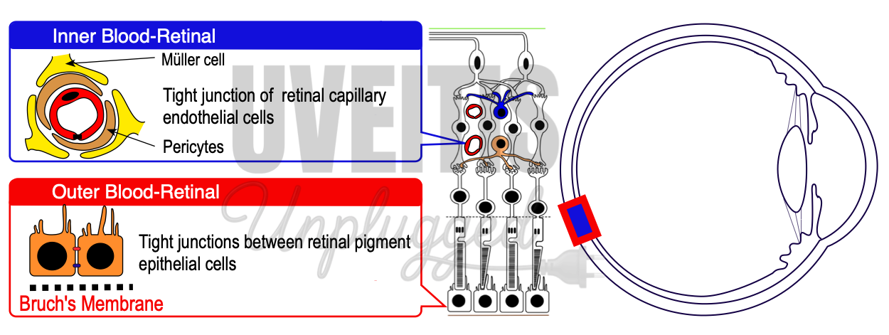
The BRB is formed by two anatomically and functionally distinct components:
1. Retinal Capillary Endothelium (Inner BRB)
Retinal capillaries consist of continuous endothelial cells connected by tight junctions that are non-leaky. These capillaries lack fenestrations, a feature shared with cerebral capillaries, which contributes to their barrier function. They are supported by a thick basement membrane, pericytes, and surrounding glial cell (notably Müller cell) processes.
Pericytes play an important role in regulating capillary tone, permeability, endothelial stability, and angiogenesis, while glial cells contribute to structural and metabolic support of the barrier.
2. Retinal Pigment Epithelium (Outer BRB)
The retinal pigment epithelium (RPE) forms the major barrier between the neural retina and the choroidal circulation. Extensive apical tight junctions between RPE cells restrict the passage of substances from the fenestrated choriocapillaris into the retina.
Although Bruch’s membrane offers only minor resistance, its negative charge limits the movement of negatively charged molecules. In addition to its barrier role, the RPE actively transports fluid from the subretinal space into the choroidal circulation, helping maintain retinal attachment and homeostasis.
The choriocapillaris itself has a fenestrated endothelium, allowing efficient delivery of oxygen and nutrients to the outer retina.
Transport Across Blood–Ocular Barriers
Substances cross blood–ocular barriers by two main mechanisms:
- Transcellular transport, which occurs through cells via passive diffusion, facilitated diffusion, or active transport
- Paracellular transport, which occurs between cells and is tightly restricted by junctional complexes
Because these barriers are impermeable to many essential water-soluble molecules such as glucose and amino acids, specialized energy-dependent transporters are required to supply nutrients to intraocular tissues.
Functional Significance and Shared Features
Both the blood–aqueous and blood–retinal barriers:
- Separate a highly regulated ocular compartment from richly vascularized tissues
- Enable controlled delivery of oxygen and nutrients and efficient removal of metabolic waste
- Maintain osmotic balance in avascular structures such as the cornea, lens, vitreous, and retina
- Allow selective passage of molecules essential for ocular function (e.g., vitamin A for photoreceptors)
- Exclude larger or potentially harmful molecules that could compromise transparency or neural activity
Reference:
- Cunha-Vaz J. The blood-ocular barriers. Surv Ophthalmol. 1979 Mar-Apr;23(5):279-96.
- Freddo TF. A contemporary concept of the blood-aqueous barrier. Prog Retin Eye Res. 2013 Jan;32:181-95.
- Cunha-Vaz J, Bernardes R, Lobo C. Blood-retinal barrier. Eur J Ophthalmol. 2011;21 Suppl 6:S3-9.