
PG Series: Classification & Clinical Features of Uveitis
A simplified version for the Ophthalmology Postgraduates

Nodules of the iris.
Keratic precipitates
Acute anterior uveitis.
Discuss diagnosis and management of acute Iridocyclitis.
What are different classifications of uveitis? Discuss briefly about the granulomatous uveitis and its management.
Classification of Uveitis:
A. Pathological or Woods Classification:
- Woods classification is also known as pathological or clinicopathological classification
- Uveitis is classified as granulomatous or nongranulomatous on the basis of the predominant clinical characteristics.
|
Granulomatous Uveitis |
Nongranulomatous Uveitis |
|
Insidious onset and chronic course |
Sudden onset and acute course |
|
Absent or mild congestion |
Severe episcleral congestion |
|
Iris nodules (Keoppe’s and Bussaca’s nodules) are common |
Iris nodules are uncommon |
|
Medium to large keratic precipitates (Mutton fat KPs) are seen |
Fine, small keratic precipitates are seen |
|
Posterior segment involvement is common |
Posterior segment involvement is uncommon |
B. IUSG classification:
- International Uveitis Study Group (IUSG) classification of uveitis was originally devised by the International Uveitis Study Group in 1987.
- This classification is largely based on the anatomic position of the inflammation within the eye
|
Anterior uveitis |
Iritis, Anterior cyclitis, Iridocyclitis |
|
Intermediate uveitis |
Pars planitis, Posterior cyclitis, Hyalitis, Basal retinochoroiditis |
|
Posterior uveitis |
Focal, multifocal, or diffuse choroiditis, chorioretinitis, retinochoroiditis, or neurouveitis |
|
Panuveitis |
C. SUN Classification:
- Most recent and widely accepted version of classifying uveitis is the Standardization of Uveitis Nomenclature (SUN) Classification.
- According to SUN classification, uveitis was divided according to their primary site of inflammation
|
Type |
Primary site of inflammation |
Includes |
|
Anterior uveitis |
Anterior chamber |
Iritis Iridocyclitis Anterior cyclitis |
|
Intermediate uveitis |
Vitreous |
Pars planitis Posterior cyclitis Hyalitis |
|
Posterior uveitis |
Retina or choroid |
Focal, multifocal, or diffuse choroiditis Chorioretinitis Retinochoroiditis Retinitis Neuroretinitis |
|
Panuveitis |
Anterior chamber, vitreous and retina or choroid |
Descriptors of Uveitis:
|
Category |
Descriptor |
Description |
|
Onset |
Sudden Insidious |
|
|
Duration |
Limited |
< 3 months duration |
|
Persistent |
> 3 months duration |
|
|
Course |
Acute |
Episode characterized by sudden onset and limited duration |
|
Recurrent |
Repeated episodes separated by periods of inactivity without treatment 3 months in duration |
|
|
Chronic |
Persistent uveitis with relapse in 3 months after discontinuing treatment |
|
Clinical features of uveitis:
Common symptoms of uveitis:
|
Type of uveitis |
Symptoms |
|
Anterior uveitis |
Pain, redness, irritation, watering, photophobia |
|
Intermediate uveitis |
Floaters, blurred vision, |
|
Posterior uveitis |
Diminution of vision, Metamorphopsia, Positive Scotoma |
|
Symptoms |
Causes |
|
Pain |
Mainly due to ciliary spasm. Ciliary body is innervated by the trigeminal nerve, therefore often the pain is radiating to jaw, periorbital region etc. |
|
Redness |
Because of the congestion and engorgement of ciliary blood vessels, also called ciliary flush or ciliary congestion. |
|
Watering |
Reflex lacrimation |
|
Photophobia |
Photophobia is … often accompanied by blepharospasm |
|
Floaters |
Manifestation of cells and debris in the vitreous cavity. |
|
Blurred vision |
Blurred vision is mostly cloudy media like vitritis |
|
Diminution of vision |
Diminution of vision usually occurs from involvement of macula and optic nerve head |
|
Metamorphopsia |
Usually occurs due to macular involvement (objects appear smaller than they are-micropsia ; sometimes larger- macropsia) |
|
Positive scotoma |
Perception of a black spot in front of the eye corresponding to a lesion |
Signs
|
Ciliary injection |
Manifest by a ring of dilated episcleral vessels radiating from the limbus |
|
Constricted pupil |
Various chemical mediators of the inflammation act as irritant on the muscles of iris & since the sphincter muscle overcomes the action dilator muscle >>constriction of pupil Hyperaemia of the radially arranged vessels of iris >> Mechanical constriction of pupil |
|
Muddy iris |
Large exudation of fluid in to tissue spaces of iris >> swelling of iris >> loss of fine, delicate pattern of iris |
|
Sluggishly reacting pupil |
Large exudation of fluid in to tissue spaces of iris >>swelling of iris >> sluggishly reacting pupil |
|
Cells |
Various inflammatory cells move across iris to the aqueous >>These cells in AC circulate due to the convection current (cooler cornea & warm iris) >> Cells |
|
Flare |
Turbidity of the aqueous due to transudation of proteins across the inflamed blood vessels |
|
Keratic Precipitates |
Desquamation of corneal endothelium due to inflammation ® Inflammatory cells floating under convection current in AC >> stick to the endothelium >> KP |
Keratic Precipitates (KPs):
KPs are cellular deposition on corneal endothelium during uveitis.
Pathogenesis of KP: Migration of inflammatory Cells across the iris and ciliary vessels to aqueous humour ® In AC, these cells come across a convection current (due to temperature difference between inflamed warm iris & relatively cooler cornea) ® They start moving AC ®Because of the altered nutrition (inflammatory products in aqueous), corneal endothelium becomes sticky and may desquamate in places. And circulatory cells due to centrifugal convection current (convection currents in the anterior chamber that rise along the warm iris and fall along the cool cornea) and gravity often sticks on the corneal endothelium, which are termed as Keratic precipitates (KP).
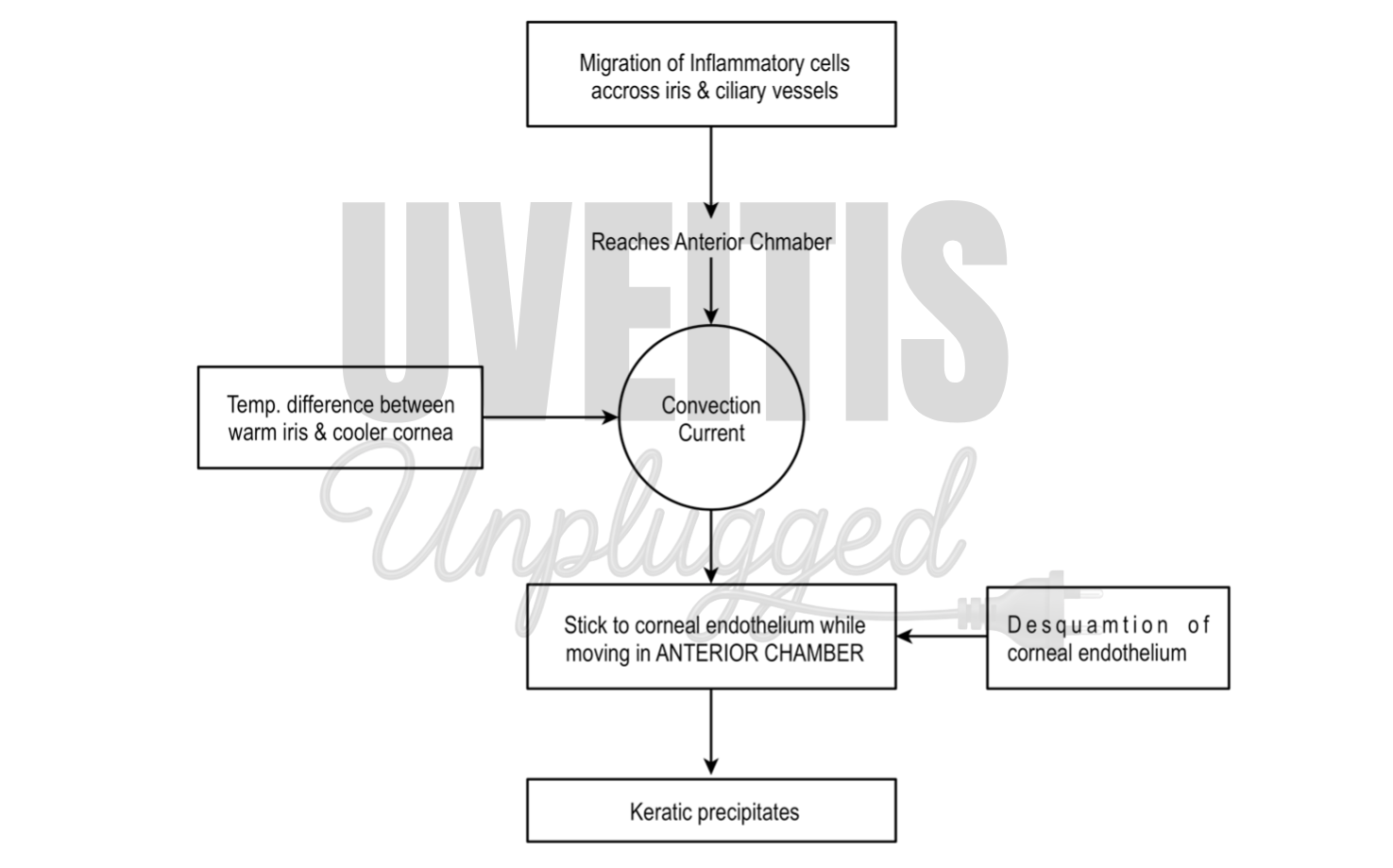
|
Distribution of KPs |
|
|
Arlt's triangle |
Base down triangular area in the lower part of the cornea |
|
Turk’s Line |
Arranged in a linear fashion (vertical line) |
|
Scattered over whole cornea |
Viral infections, and Fuchs' heterochromic iridocyclitis |
|
Type of cells |
|
|
Acute inflammation |
Polymorphonuclear leucocytes |
|
Chronic inflammation |
Lymphocytes Plasma cells and pigment cells |
|
Morphology of KPs |
|
|
Fresh KP |
White and round |
|
Old KP |
Pigmented, faded, irregular crenated margins |
|
Types of KPs |
|
|
Mutton fat KPs |
Large KPs, clusters of macrophages and epithelioid cells, pathognomonic of granulomatous inflammation. |
|
Fine KPs |
Viral infections, Nongranulomatous uveitis |
|
Stellate KPs |
Fuchs' heterochromic iridocyclitis |
Cells
- Cells in the aqueous is usually seen when there is inflammation of iris and ciliary body.
- Cells in the anterior chamber are counted using slit-lamp with a beam of 1 x 1 mm slit and graded according the SUN classification
|
Grade |
Cells in Field of 1 mm by 1 mm slit beam. |
|
0 |
< 1 |
|
0.5+ |
1–5 |
|
1+ |
6–15 |
|
2+ |
16–25 |
|
3+ |
26–50 |
|
4+ |
> 50 |
Inflammatory anterior chamber cells are generally white in colour and should be not be confused with pigmented cells. Pigmented cells can be iris pigments, dead erythrocytes, or macrophages filled with pigment like melanin.
Iris pigments can be seen in the anterior chamber after dilatation and should be distinguished from cells.
Flare: Aqueous humor is considered optically empty and if a slit- lamp beam is passed through it, the beam cannot be seen. In case of inflammation, when breakdown of the blood-aqueous barrier occurs, there is increased protein content in the aqueous and if slit beam is obliquely aimed across the anterior chamber, the path of the beam can be seen, which is termed flare.
Flare is graded according to the scheme proposed by SUN classification
|
Grade |
Description |
|
0 |
None |
|
1+ |
Faint |
|
2+ |
Moderate (iris and lens details clear) |
|
3+ |
Marked (iris and lens details hazy) |
|
4+ |
Intense (fibrin or plastic aqueous) |
Flare is often the first sign of uveitis and it may persist despite adequate control of inflammation.
Flare can also be measured using a laser flare photometry, which quantifies anterior chamber protein by measuring light scattering of a helium-neon laser beam in the anterior chamber
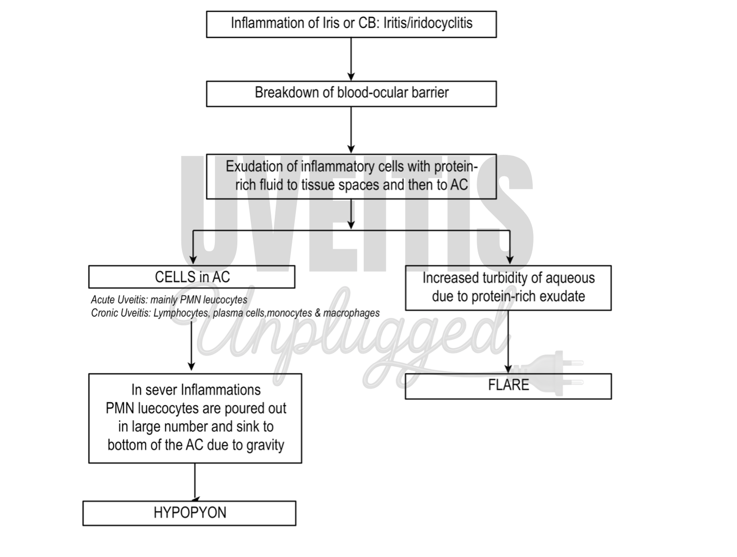
Iris nodules:
- Iris nodules are very important signs of systemic disease. Iris nodules can be seen in both inflammatory & no-inflammatory conditions.
- Koeppe & Busacca nodules are pathognomic of granulomatous uveitis.
|
Name |
Location |
Condition |
|
Koeppe nodules |
Pupillary border |
Granulomatous Uveitis |
|
Busacca nodules |
Anterior surface of the iris |
Granulomatous Uveitis |
|
Berlins nodules |
Angle of anterior chamber |
Sarcoidosis |
|
Lisch nodules |
Surface of the iris |
Neurofibromatosis type-I, not related to uveitis |
Examination of vitreous:
- Cells in the anterior vitreous or retrolental space should be looked for after pupillary dilation. Though there is no standard grading system for vitreous cells, documentation of this finding is important for follow-up of a uveitic case.
- Vitreous should be carefully examined with a 78/90D and also with indirect ophthalmoscope with indentation for snow ball opacities, snow-banking in pars plana region and vitreous strands
- For classifying vitreous haze, SUN classification has adopted The National Eye Institute system for grading vitreous haze with the proviso that the designation “trace” be recorded as 0.5+.
|
0 |
nil |
|
0.5+ |
trace |
|
1+ |
Few opacities, mild blurring |
|
2+ |
Significant blurring, but still visible |
|
3+ |
Optic nerve visible, no vessels visible |
|
4+ |
Dense opacity obscures optic nerve head |
Examination of Fundus:
- Optic disc should be carefully examined with the help of slit lamp biomicroscopy. Disc hyperaemia, disc oedema, or optic neuritis is seen in various uveitic conditions. Glaucomatous damage to the optic disc due to Secondary glaucoma, Neovascularization of the optic disc, optic disc granuloma and optic atrophy may also occur in uveitic patients.
- Examination of retinal vasculature for vasculitis, vascular sheathing, and accumulation of inflammatory cells around vessels is important. Vascular sheathing is seen as white parallel lines along vessels. Sometimes inflammatory exudates are seen around the vessels in patients with sarcoidosis, known as candle-wax drippings. Also it is important to determine whether retinal veins, retinal arteries, or both are affected as it can help in differential diagnosis of the uveitic entity.
|
Arterial involvement (arteritis) |
Venous involvement (phlebitis) |
|
|
- Careful examination of the posterior segment can reveal inflammatory patches in the fundus. It is important to distinguish such lesions whether it involves retina or choroid or both. Sometimes these lesions are associated with subretinal fluid or localized haze in vitreous.
|
Retinitis |
Choroiditis |
|
|
- Often a patient can present with a retinal detachment. Exudative retinal detachments can be seen in a number of ocular inflammatory diseases like Vogt–Koyanagi–Harada syndrome. One should be able to distinguish rhegmatogenous retinal detachment from such cases, which requires a surgical management. Sequlae to vasculitis can lead to development of traction retinal detachment and should be dealt properly.
- Meticulous examination of the fovea with slit lamp biomicroscopy often helps to identify cystoid macular oedema, choroidal neovascular membrane or sight threatening inflammatory lesions like serpiginous choroiditis. Cystoid macular oedema is a common in patients with uveitis which if long standing can lead to formation of macular hole.