
PG Series: Ocular Toxoplasmosis

Toxoplasmosis is caused by Toxoplasma gondii, an obligate intracellular protozoan.
- Life cycle of parasite is completed or require following hosts:
- Definitive host (An organism which supports the adult or sexually reproductive form of a parasite) = Cat
- Intermediate hosts (An organism that supports the immature or non-reproductive form of a parasite) = Mice, Pig, Cow and Humans.
- There are primarily three strains of Toxoplasma gondii – namely type I,II and III. Many atypical strains of the organism are also reported.
- The parasite exists in the following forms:
|
Sporozoites |
|
contained within an oocyst (sporocyst) |
|
Bradyzoites |
Inactive and are that most commonly develop in the brain, eye, heart, skeletal muscles and lymph nodes. They may lie dormant for many years without provoking an inflammatory reaction |
contained within tissue cysts |
|
Tachyzoites |
Proliferating active form responsible for tissue destruction and inflammation |
Released following rupture of the wall of a cell containing bradyzoites. |
Lifecycle: Sexual reproduction of the parasite occurs only in the cells of the feline intestinal tract. The products of the sexual reproduction, oocysts contain sporozoites. Once oocysts are shed from cats, they can remain viable in the environment for long periods of time. Following ingestion, oocysts develop into the tachyzoites. Tachyzoites eventually undergo asexual reproduction and produce bradyzoites, which persist in the tissues in a viable cyst form. This asexual reproduction can occur in either cats or in intermediate hosts like pigs, cows, human beings. The cysts contain hundreds to thousands of bradyzoites, and have a propensity for cardiac tissue, muscle, and neural tissue, including the retina.
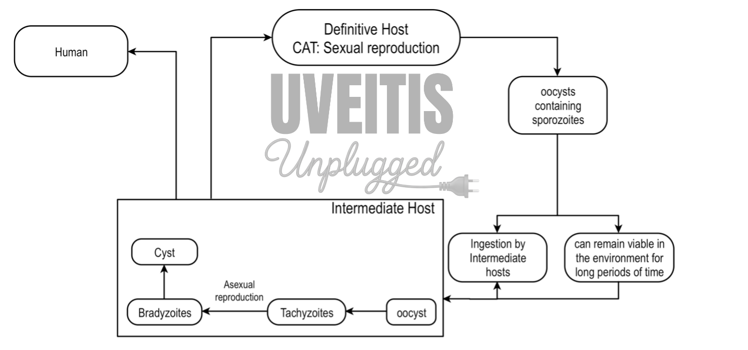
In human being two forms of the parasite are found – cysts and tachyzoites. Toxoplasma gondii believed to invade human retina and can transform into cystic form in inner sensory retina. Necrotizing retinitis occurs when the cyst ruptures and tachyzoites replicate in retinal cells with inflammatory reaction to surrounding retina and choroid.
In contrast to tachyzoites, which cause tissue destructive inflammatory disease as they proliferate, cysts do not stimulate tissue inflammation. Cysts have been found to remain viable outside the host in soil for at least 1 year.
Mode of human infection:
- Ingestion of undercooked meat (lamb, pork, beef) containing bradyzoites of an intermediate host.
- Ingestion of sporocyst following inadvertent contamination of hands when disposing of cat litter trays and then subsequent transfer on to food. Infants may also become infected by eating dirt, soil (pica) containing sporocyst.
- Ingestion of contaminated water, fruit, or vegetables with oocysts
- Transplacental spread of the parasite (tachyzoite) can occur if a pregnant woman becomes infected.
- Blood transfusion or organ transplantation (rare)
Clinical features
- Presentation: unilateral sudden onset of floaters, visual loss and photophobia.
- ‘Spill-over’ anterior uveitis, usually granulomatous
- Solitary inflammatory focus near an old pigmented scar (‘satellite lesion’)
- Retinochoroiditis:
- MC manifestation of OT.
- Starts as yellowish-white lesion involving inner retina with adjacent retinal oedema® gradually progresses to involve full thickness of retina and choroid
- Active retinochoroiditis in the midst of severe vitritis may give characteristic ‘Headlight in the fog’ appearance
- Recurrence is common and can occur
- at the margin or closer to a pre-existent retinochoroidal scar (satellite lesion)
- Isolated focal lesion
- Kyrieleis arteritis/plaques:
- Clinically manifested as segmental retinal periarteritis.
- Affects arteries and do not leak dye in FFA
- Not specific and can be seen in various other conditions - Rickettsia conorii, Mycobacterium tuberculosis, Treponema pallidum, and varicella-zoster virus (VZV) infections.
- Atypical features in immunocompromised patients :
- Large, multiple, and/or bilateral lesions
- Neuroretinitis, Punctate outer retinal toxoplasmosis (PORT), unilateral pigmentary retinopathy simulating retinitis pigmentosa, and other forms of intraocular inflammation in the absence of retinochoroiditis.
- PORT is characterized by small, multifocal lesions at the level of the outer retina, with exudation to subretinal space and scant overlying vitreal inflammation
- Extensive confluent areas of retinitis mimicking viral retinitis.
Congenital toxoplasmosis
Toxoplasmosis is transmitted to the fetus through the placenta when a pregnant woman becomes infected.
Severity of involvement of the fetus is dependent on the duration of gestation at the time of maternal infection.
Clinical Manifestations:
- Most cases of congenital systemic toxoplasmosis are subclinical and bilateral healed chorioretinal scars are usually discovered later in life, either by chance or when the child is found to have defective vision.
- The classic presentation of congenital toxoplasmosis is described by Sabin's tetrad and it includes
-
- Retinochoroiditis,
- Hydrocephalus or microcephaly,
- Intracranial calcifications, and
- Cognitive impairment
-
- Retinochoroidal lesion is the MC abnormality in congenital toxoplasmosis;
-
- Bilateral
- Has a predilection for the posterior pole and macula
-
- Infections occurring towards the end of the second trimester usually result in disease that can be detected at birth such as macular scars (Fig. 11.23B), while those occurring later in the third trimester may result in normal examination at birth, but the appearance of ocular or neurological symptoms in the future.
|
Systemic manifestation |
Ocular manifestation |
|
Diarrhea, Vomiting Hydrocephalous Macro/microcephaly Jaundice Lymphadenopathy Low birth weight Swollen Spleen and Liver |
Retinochoroiditis Bilateral chorioretinal scars Microcornea Microphthalmos Nystagmus Strabismus |
Laboratory Investigations Ocular Toxoplasmosis:
- Diagnosis of OT is almost always clinical
- Serologic evaluation (by indirect fluorescent antibody and ELISA methods) is aimed at detection of specific anti-T. gondii antibodies which indirectly confirm the exposure to the parasite.
|
Anti-T. gondii antibodies |
Appears |
Remains |
Crosses Placenta |
Clinical Significance |
|
IgG antibodies |
after the first 2 weeks of infection |
detectable for life |
Yes |
Negative antibody titer essentially rules out the diagnosis |
|
IgM antibodies |
early during the acute phase of infection |
detectable for less than 1-2 year |
No |
In newborns confirms congenital infection and in adults indicative of acquired disease |
|
IgA antibodies |
variable |
disappear by 7 months |
No |
Useful in a diagnosis of congenital toxoplasmosis in a fetus or newborn (during this period, IgM production is often weak and the presence of IgG antibodies may indicate passive transfer of maternal antibodies in utero.) |
- Intraocular antibody production was determined by calculation of the Goldmann-Wittmer (GW) coefficient, which is the quotient of the relative amounts of antitoxoplasma antibodies in the eye and serum and is calculated as follows: antibody titer ocular fluid / total IgG ocular fluid: antibody titer serum / total IgG serum
- A Goldmann-Wittmer coefficient of greater than 3 is considered as evidence for T. gondii–specific antibody production in the eye with a test specificity of 100% and a sensitivity of 74%
Treatment:
- OT may be self-limited and the decision to treat depends on- 1. Location, size of the lesion and 2. Immune status of the patient
- The aim of the treatment is 1. to arrest parasite multiplication during the active period of retinochoroiditis and 2. to minimize damage to the retina and optic disc
- Most current medications are effective against tachyzoites, but not against tissue cysts
|
Classic regimen: Triple therapy
|
Leukopenia and/or Thrombocytopenia Folinic acid (15 mg every day) is added to prevent myelosuppression |
|
Oral Clindamycin (300 mg orally four times daily) |
Pseudomembranous colitis |
|
Usually well tolerated, although sulfonamide-related reactions may occur |
|
Azithromycin (250 mg/day) |
Diarrhea, loose stools |
|
Atovaquone (750 mg three/four times daily orally) |
|
|
Intravitreal Clindamycin 1.5 mg |
Found to be non-toxic to the retina |
Corticosteroid:
- Administration of corticosteroids without antiparasitic medication might result in exacerbated ocular disease with enormous tissue destruction and higher numbers of tissue cysts.
- Oral corticosteroids are usually administered either at the time of antimicrobial therapy or within 48 hours
- Topical corticosteroids are used to treat prominent anterior segment inflammation.