
PG Series: Granulomatous uveitis

Questions Asked in the Exam:
Discuss briefly about the granulomatous uveitis and its management.
Ocular manifestations in leprosy
Ocular manifestations of TB
How you will investigate a case of chronic uveitis?
A granulomatous uveitis is characterized by following features
- Granulomatous Uveitis
- Insidious onset and chronic course
- Absent or mild congestion
- Iris nodules (Keoppe’s and Bussaca’s nodules)
- Medium to large keratic precipitates (Mutton fat KPs)
- Posterior segment involvement
Causes of granulomatous uveitis can be summarised as:
|
Infective |
Noninfective |
|
Tuberculosis Leprosy Syphilis Viral Lyme disease Borrelia |
Sarcoidosis Vogt-Koyanagi-Harada disease Sympathetic Ophthalmia |
Ocular Tuberculosis:
Tuberculosis (TB) is a chronic granulomatous infection caused by human tubercle bacilli (Mycobacterium tuberculosis) or bovine (Mycobacterium bovis)
Ocular involvement occurs in about 1 % to 2% of patients with TB
Tuberculous uveitis is a chronic, granulomatous infection that can involve any part of the uveal tract
Primary ocular TB = eye is the primary portal of entry
Secondary ocular TB = occurs by haematogenous dissemination or by contiguous spread from adjacent structures.
Clinical features
Ocular Manifestations of Tuberculosis: Ocular TB may have protean manifestations, & may involve any part of the eye, ocular adnexae or orbit
|
Eyelids |
Rare, may present as localized nodule simulating chalazion |
|
Lacrimal gland & orbit |
Localized mass or granuloma |
|
Conjunctiva |
Conjunctival ulceration, granuloma and hypertrophied papillary lesion |
|
Phlycten |
Delayed hypersensitivity reaction in cornea or conjunctiva to mycobacterial antigen Localized elevated nodule at limbus or at conjunctiva with leash of blood vessels |
|
Sclera |
Diffuse and nodular scleritis Nodular Episcleritis |
|
Cornea |
Interstitial keratitis Disciform Keratitis |
|
Anterior Uveitis |
Granulomatous anterior uveitis Large MF KPs Iris nodules- Koeppe & Busacca nodule Broad based posterior synechiae Anterior chamber granuloma (rare) |
Anterior Segment manifestations of Ocular Tuberculosis
|
Intermediate Uveitis |
Low grade, smouldering, chronic uveitis with pars plana exudates, snowball opacities, snow banking, peripheral vascular involvement, peripheral retinochoroidal granuloma |
|
Choroidal Tubercle |
Post pole Multiple lesions > Solitary lesion 0.3-3 mm in diameter Yellowish white in colour |
|
Choroidal Tuberculoma |
Solitary lesion Yellowish-white raised lesion with indistinct margin 2-3 DD or larger |
|
Subretinal Abscess |
Large yellowish subretinal mass with exudative retinal detachment USG: Hyperechoic lesion with internal mixed echogenicity |
|
Tubercular Serpiginous like choroiditis |
Usually start as multifocal choroiditis to progress and become confluent Associated with significant vitreous inflammation |
|
Panuveitis/Endophthalmitis |
Rare Can result from breaking up of SR abscess into vitreous cavity |
|
Retinal Vasculitis |
Characterised by perivascular cuffing, retinal haemorrhages with peripheral retinal capillary closure and neovascularisation Eales disease-like picture in patients with Indian subcontinent |
|
Tuberculous Neuroretinitis |
Retrobulbar optic neuritis Macular star & optic disc oedema Association with tubercular meningitis |
Posterior Segment manifestations of Ocular Tuberculosis
Diagnosis
The diagnosis of ocular TB is usually difficult to make.
|
Definitive diagnosis |
Presumptive diagnosis |
|
Demonstration of the bacilli in eye |
Demonstration of the bacilli in other tissues |
|
Provides direct evidence of the infection |
Provides indirect evidence of the infection |
|
Tissue samples are most difficult to obtain |
Relatively easy to obtain |
Laboratory Diagnosis of Ocular Tuberculosis:
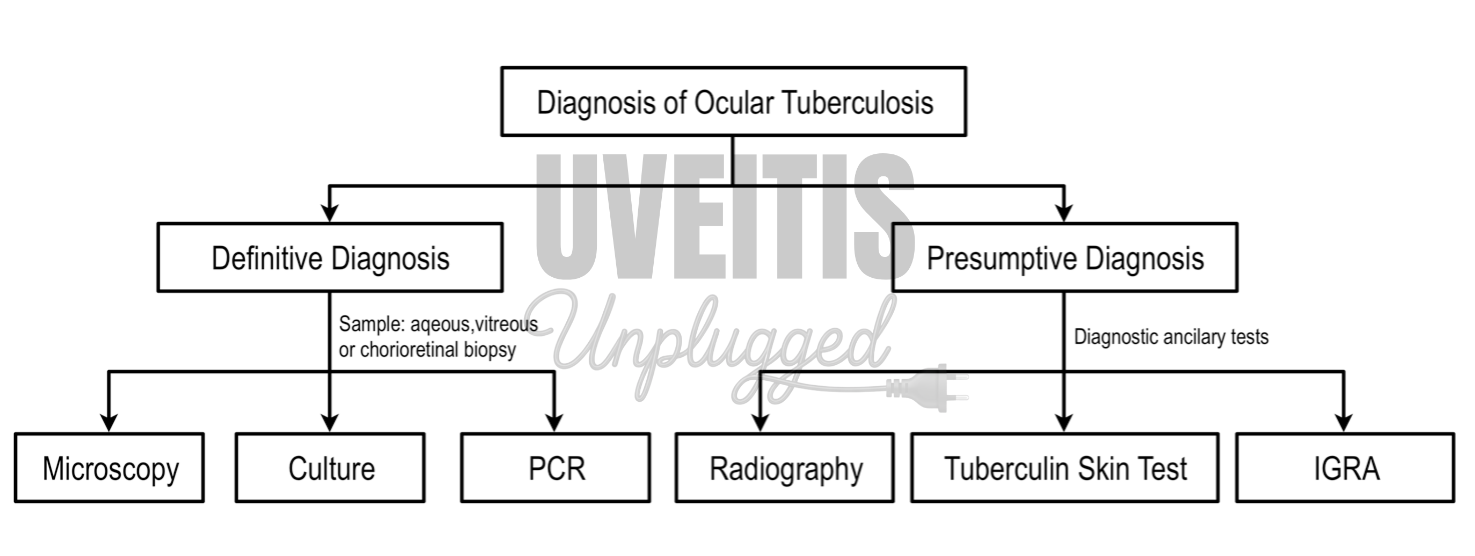
Definitive Diagnosis:
|
Name of the test |
Features |
|
Microscopy |
The sample is centrifuged and then stained either with Ziehl Neelsen stain or special immunofluorescent stains. |
|
Cultures |
Demonstration of acid-fast bacilli on direct smear and culture from intraocular fluid or tissue sample is diagnostic |
|
Histopathology & PCR of tissue sample |
Aqueous or vitreous aspirate can be collected for microbiological analysis Chorioretinal, iris, or vitreous biopsy may be used to obtain tissue for histopathologic analysis and for PCR analysis for mycobacterial genome |
|
Polymerase chain reaction (PCR) |
PCR for the detection of M. tuberculosis DNA : Multi-target PCR using three genes: protein b, MPB 64 and IS6110 Real-time/ or q-PCR Nested PCR |
While about 5000 to 10,000 bacilli per ml must be present to be detected on stained smears, 10-100 viable bacteria per ml may suffice to be grown in culture.
Presumptive Diagnosis:
|
Name of the test |
Features |
|
Sputum Examination |
The WHO recommends sputum microscopic examination to establish the diagnosis of PTB because it is highly specific, inexpensive, and widely established. However, at least 106 organism/ml of sputum are required for detection in smear and it is rarely performed for the diagnosis of ocular tuberculosis |
|
Tuberculin Skin Test (TST) |
A positive TST in a patient with uveitis does not mean that the uveitis is caused by TB |
|
Interferon Gamma Release Assay (IGRA) |
It measures Interferon-g released by the sensitized T cells after stimulation by the Mycobacterial tuberculosis antigens. Though highly specific, it does not differentiate between an active or latent infection. The sensitivity of IGRA is similar to TST but being more specific, it helps in evaluating a positive TST and to exclude a false-positive one. |
|
Chest radiography: CXR/ HRCT Chest |
To detect signs of active pulmonary tuberculosis like consolidation, cavitation, satellite lesions or hilar lymphadenopathy or signs of old healed TB including fibrosis, calcified pulmonary nodules or hilar lymph nodes. |
|
Multidrug Gene X |
a cartridge based nucleic acid amplification test, automated diagnostic test that can identify Mycobacterium tuberculosis (MTB) DNA and resistance to rifampicin (RIF) by nucleic acid amplification test (NAAT). |
|
Isoniazid therapeutic trials |
The patient is administered isoniazid at 300 mg/day and examined every week. If ocular inflammation improves after 1 to 2 weeks of therapy, the pt. is considered to have a positive test response. |
Tuberculin Skin Test:
According to CDC guidelines, the test is reported positive
- ≥15 mm in non-endemic population,
- ≥10 mm in TB endemic population
- ≥5 mm in immune deficient patients, those having chest radiographic signs of healed TB and those having recent contact with active TB
False negative test = Erroneous testing or anergy conditions like sarcoidosis, uraemia, Hodgkin’s disease etc.
False positive test = Prior exposure to atypical mycobacteria or BCG vaccination (effect of BCG vaccine usually wane away by 3-5 years).
Booster effect = Increased reaction seen on sequential PPD testing and may occur either due to a remote tuberculosis infection or by sensitization with atypical mycobacteria.
Limitation: 1. Cross-reactivity of the antigen used in TST with BCG and environmental nontuberculous bacteria. 2.Booster-effect on repeated injections (false-positive result) 3. Immunocompromised patients (false-negative result) 4. Cannot differentiate between latent infection and active disease
Differential diagnosis of tuberculous uveitis:
- Sarcoidosis
- Syphilitic gummas
- Toxoplasmosis
- Toxocariasis
- Candidiasis
- Coccidiomycosis
- Nocardiosis
- Brucellosis
- Behçet's syndrome
- Cat-scratch disease
- Leprosy
- Lyme disease
- Leptospirosis
- Chronic granulomatous disease
- Tumor metastasis
- Subretinal cysticercosis
Treatment:
CDC recommendations Use of all four drugs (isoniazid, rifampicin, pyrazinamide and ethambutol)for an initial 2-month period followed by a choice of different options over next 4 to 7 months for the treatment of TB
Antitubercular Treatment:
Two months of 4-drug therapy.
- Isoniazid 5mg/Kg daily
- Rifampicin 450mg daily
- Pyrazinamide 30mg/Kg daily
- Ethambutol 15mg/Kg daily
Followed by 4-month continuation phase of Isoniazid & Rifampicin. Many authors advocate the use of ATT for 9 months
Leprosy
- Leprosy (Hansen's disease) is a chronic, granulomatous disease caused by the intracellular acid-fast bacillus Mycobacterium leprae.
- Common in Indian subcontinent, sub-Saharan Africa, and Southeast Asia.
- Humans are the only natural host for M. leprae
- M. leprae has a tropism for body areas that have low temperatures, particularly the skin, peripheral nerves, nasal mucosa, and anterior segment of the eye.
-
Leprosy was the first documented bacterial infection in humans.
Leprosy has the highest incidence of ocular complications of any systemic disease.
The mode of transmission: through the mucous membrane of the upper respiratory tract or the skin.
Leprosy is often divided into two major subtypes: tuberculoid leprosy and lepromatous leprosy.
|
Tuberculoid leprosy |
Lepromatous leprosy |
|
Organism induces a strong cell mediated immune response, |
In these patients little immunity follows the infection |
|
few organisms are thus found invading organ tissues |
a plethora of organisms are found throughout the body |
|
with lepromin is highly positive (Mitsuda reaction) |
. Intraocular inflammatory disease presenting as acute reactional states (Type I and Type II) that arise from acute changes in the immune status occur more commonly in this .They are composed predominantly of macrophages with numerous acid-fast bacilli. Iris pearls consist of macrophages filled with bacilli. |
|
Uveitis is more common |
Uveitis: less common |
Ocular manifestations of leprosy:
|
Lid & Facial Muscles |
Madarosis, Ectropion, Entropion, Trichiasis, Ptosis Orbicularis oculi weakness, Lagophthalmos, Facial paralysis and anaesthesia |
|
Conjunctiva |
Pterygium, Conjunctivitis |
|
Sclera |
Episcleritis/scleritis |
|
Cornea |
Prominent corneal nerves, Decreased corneal sensitivity, Corneal anesthesia, Exposure keratitis |
|
Uveal tract |
Pinpoint pupils, Chronic granulomatous uveitis, Iris pearls, Iris atrophy |
|
Lens |
Cataract |
Uveitis :
- Usually asymptomatic, with minimal or no symptoms until late in the disease process
- Minimal ciliary or conjunctival congestion
- Bilateral chronic granulomatous anterior uveitis – MF KPs or few, scattered, fine KPs, few anterior chamber cells, mild-to-moderate flare
- Iris pearls- small, white glistening substances on the anterior surface of the iris or at the pupillary border. These 'pearls' slowly enlarge and coalesce. become pedunculated and drop into the inferior angle, where they may be observed by gonioscopy from which they eventually disappear
- Iris becomes atrophic and the pupil miosed
- Posterior synechiae are uncommon
Complications:
- Pinpoint pupils,
- Glaucoma or
- Iris atrophy,
- Ciliary body damage and Hypotony
- Complicated cataract
Diagnosis:
Diagnosis usually is made easily on the basis of
- Thorough clinical evaluation
- Histology of the skin lesions or skin biopsy is used for confirmation. Iris pearls are characteristic.
- Skin tests with lepromin may be highly positive (Mitsuda reaction) in the tuberculoid type
- Culture: Attempts to culture the organism have not been successful (doubling time for the organisms is extraordinarily long, about 20 days)
Differential diagnosis :
- Tuberculosis
- Sarcoidosis
- Idiopathic anterior uveitis
- Syphilis
Treatment:
- Early screening for ocular complications: Because of the chronicity and insidious nature of this anterior uveitis
- Once detected, regular, (every 1-6 months) eye examinations are advisable, depending on the severity of the uveitis and its response to therapy
- Topical corticosteroid and mydriatic therapy along with appropriate antimicrobial therapy is mainstay of the therapy
- Dapsone, rifampin, and clofazimine are the primary antileprosy agents used. Multidrug treatment regimens are advisable because of dapsone-resistant strains of M. leprae.
- Surgical management of eyelid deformities where indicated
- When intraocular surgery is planned: Quiet eye for at least 3-month