Ocular Immune Privilege & Defense
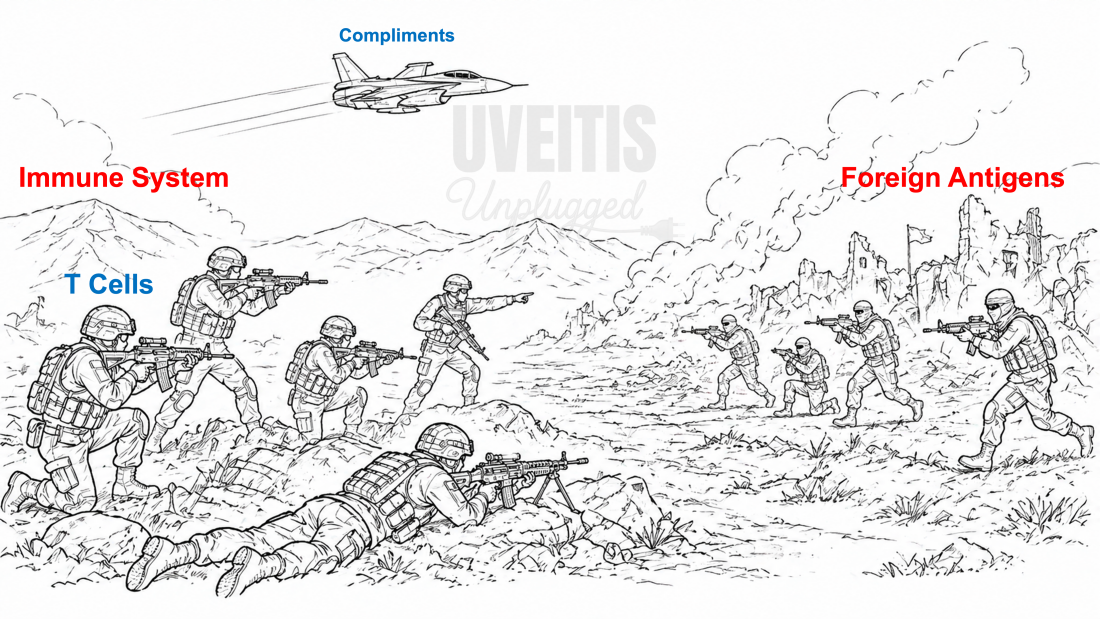
Think of the immune system as an army whose primary role is to defend the body against invading enemies. In most tissues, when a foreign antigen enters, the immune system launches a vigorous attack, deploying T cells, activating the complement cascade, and generating inflammation to eliminate the threat. While this response is highly effective against infections, it can also cause significant collateral damage to the surrounding tissue. For most organs, such damage can be repaired to some extent. The eye, however, contains several highly specialized cells that have little or no capacity for regeneration. Photoreceptors, once lost, are gone forever; corneal endothelial cells gradually decline throughout life and do not regenerate; and damage to trabecular meshwork cells can impair aqueous outflow and lead to glaucoma. Recognizing the vulnerability of these critical structures, the eye has evolved unique mechanisms that selectively suppress and regulate immune responses within its tissues. This delicate balance allows protection against pathogens while minimizing inflammatory damage, a phenomenon known as ocular immune privilege.
Immune privilege does NOT mean the eye is unprotected. It means the eye controls HOW it responds — suppressing the most destructive pathways while preserving others.
Historical Background
Our understanding of ocular immune privilege evolved through a series of landmark discoveries. In 1948, Peter Medawar demonstrated that skin grafts placed within the anterior chamber of rabbit eyes survived far longer than similar grafts placed elsewhere in the body. This observation led him to describe the eye as an “immunologically privileged site.” Several decades later, during the 1970s–1990s, J. Wayne Streilein expanded this concept by showing that antigens introduced into the anterior chamber do not elicit a conventional immune response. Instead, they induce a state of systemic immune tolerance, a phenomenon he termed Anterior Chamber-Associated Immune Deviation (ACAID). These pioneering studies established Streilein as the father of modern ocular immunology. Subsequent research in the 1990s and 2000s elucidated the molecular basis of ocular immune privilege, identifying key immunoregulatory factors such as Fas ligand (FasL), transforming growth factor-beta2 (TGF-β2), alpha-melanocyte-stimulating hormone (α-MSH), PD-L1, and complement regulatory proteins, all of which contribute to maintaining the eye’s unique immunosuppressive microenvironment.
Components of Ocular Immune Privilege
Ocular immune privilege is maintained through four major components that work together to protect the eye from excessive immune-mediated damage. The first component is the presence of specialized structural barriers, namely the blood-ocular barriers, which restrict the entry of immune cells, antibodies, and inflammatory proteins into ocular tissues. The second component is the immunosuppressive microenvironment of the eye, particularly the aqueous humor, which contains a variety of immunomodulatory factors that suppress the activation and function of immune cells. The third component involves the expression of specialized surface molecules by ocular cells, such as Fas ligand and PD-L1, which can silence, deactivate, or induce apoptosis of infiltrating immune cells. The fourth component is anterior chamber-associated immune deviation (ACAID), a unique mechanism through which antigens introduced into the eye promote systemic immune tolerance rather than a conventional inflammatory response, effectively “educating” the immune system to tolerate ocular antigens. Together, these complementary mechanisms preserve vision by minimizing inflammatory injury while maintaining the ability to respond to potential threats.
Blood-ocular Barriers
Kindly refer to the section Blood Ocular Barrier
The Immunosuppressive Cocktail
The aqueous humor, produced by the ciliary processes at a rate of approximately 2 µL per minute, is a highly specialized immunosuppressive fluid that plays a central role in maintaining ocular immune privilege. Rather than being merely a nutritive medium, it contains a remarkable array of molecules that actively suppress inflammation and promote immune tolerance.
• TGF-β2 (Transforming Growth Factor-beta2): The most abundant immunosuppressive factor in the aqueous humor. It suppresses T-cell activation, promotes the generation of regulatory T cells (Tregs), and inhibits natural killer (NK) cell cytotoxicity. It can be regarded as the eye’s principal “calm down” signal.
• α-MSH (Alpha-Melanocyte-Stimulating Hormone): Produced by the ciliary body and iris, α-MSH converts pro-inflammatory effector T cells into regulatory T cells through melanocortin receptor signaling, thereby fostering immune tolerance.
• VIP and CGRP (Vasoactive Intestinal Peptide and Calcitonin Gene-Related Peptide): These neuropeptides are released from nerves within the ciliary body and suppress both T-cell activation and the pro-inflammatory activity of macrophages.
• Thrombospondin-1 (TSP-1): Activates latent TGF-β, enhancing its biological activity. In addition, it directly suppresses NK cells and macrophages, further strengthening the eye’s immunoregulatory environment.
• Other Immunomodulatory Factors: Molecules such as macrophage migration inhibitory factor (MIF), somatostatin, and free cortisol provide additional layers of immune suppression, acting on different cellular targets but ultimately serving the common goal of limiting inflammation within the eye.
Together, these immunosuppressive factors create a unique microenvironment that restrains excessive immune responses while preserving the delicate ocular tissues essential for vision.
The ciliary body is richly innervated by the nasociliary nerve (V1 branch) carrying sympathetic, parasympathetic, and sensory fibres. These nerves ALSO release VIP and CGRP into the ciliary body, contributing directly to AH immunosuppression. This is why trauma or surgery damaging the ciliary body can disrupt immune privilege.
Clinical Perspective: Vitrectomy removes the vitreous — which also has immunosuppressive factors (similar to AH). Post-vitrectomy, the immune privilege of the posterior segment is reduced. This is one reason why intravitreal steroid implants (Ozurdex, Iluvien) are particularly useful in post-vitrectomy eyes with uveitis.
Alymphatic Nature Of the Cornea and Anterior Segment:
The healthy central cornea has NO blood vessels and NO lymphatic vessels. Lymphatics stop at the limbus. This is anatomically critical — lymphatic vessels carry antigens to regional lymph nodes (in this case, submandibular), where T cells would be activated in a normal (immunogenic) way. Without lymphatics, antigen from the AC cannot reach the lymph node via the conventional DC-lymph node-T cell priming route. Instead, it must exit via the aqueous → trabecular meshwork → bloodstream → spleen, where it triggers ACAID (tolerogenic, not immunogenic).
Surface Molecules — The Eye's Bouncers
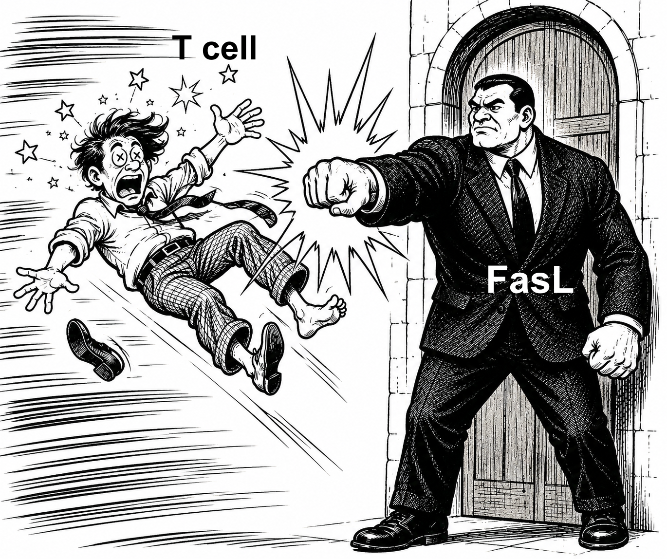
FasL (Cd95l) — The Apoptosis Trigger: Fas (CD95) is a surface protein on activated T cells. When Fas binds its ligand FasL (CD95L), it triggers apoptosis (programmed cell death) inside the T cell within hours. Here is the key fact: the corneal endothelium, iris, ciliary body, and RPE all express FasL constitutively (always, not just during inflammation). So when an activated Fas+ T cell tries to enter the eye, the FasL on the eye's surface kills it immediately. FasL is like a bouncer at the door who does not just say "no entry" — he eliminates the troublemaker on the spot. The T cell comes in, touches the corneal endothelium, and dies.
Clinical Perspective: (1) The corneal endothelium is a single layer of hexagonal cells on the posterior surface of the cornea. These cells maintain corneal clarity by pumping water out. They express FasL. This is one reason the cornea can survive across HLA barriers — donor FasL kills recipient anti-donor T cells trying to infiltrate. (2) This explains the success of penetrating keratoplasty without HLA matching. FasL + avascularity + alymphatic nature = triple protection. When any one is compromised (herpes stromal keratitis, alkali burn, repeat graft) rejection risk rises sharply. (3) Herpes simplex virus can downregulate FasL expression on corneal cells as a survival mechanism — one reason HSK is so destructive and recurrent.
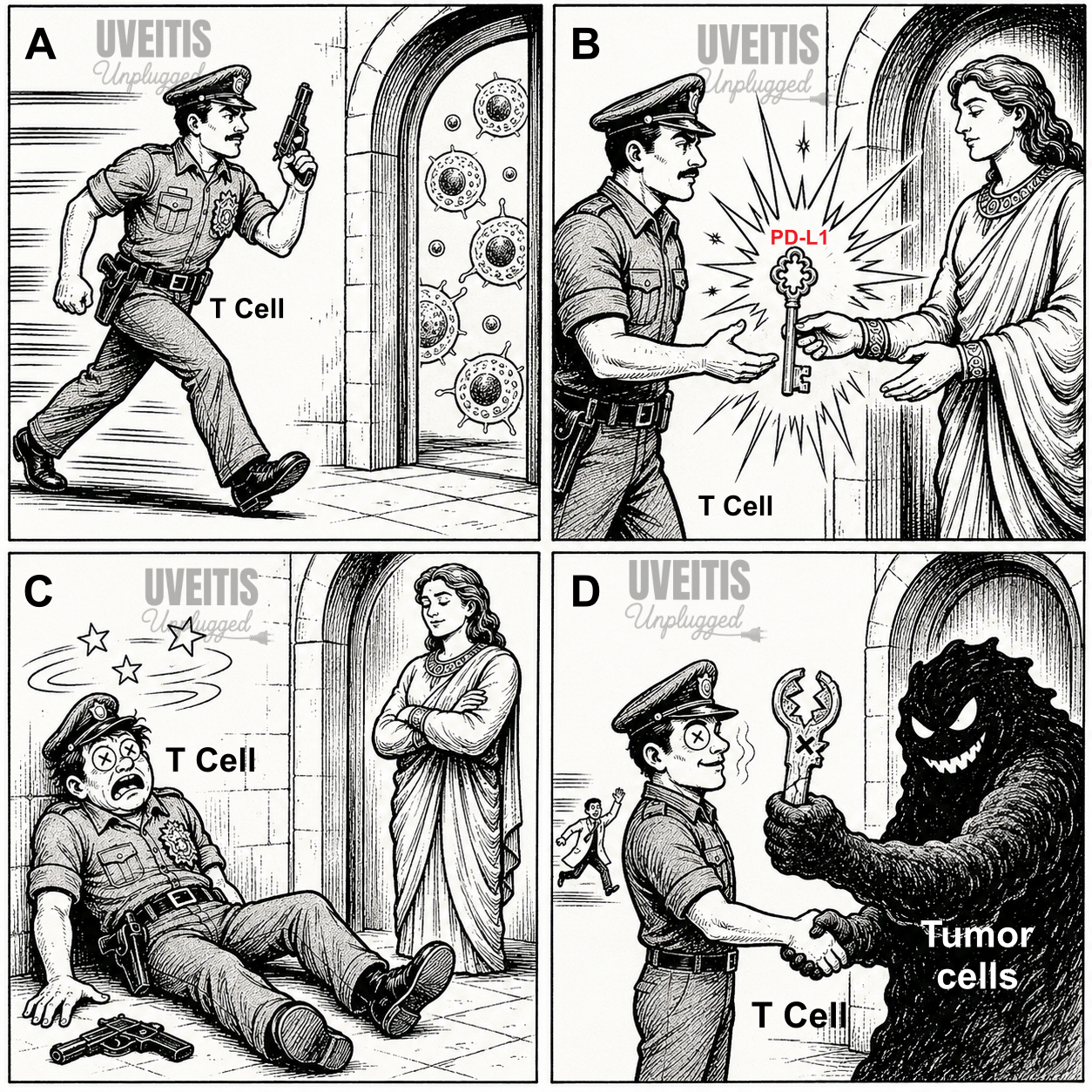
PD-L1 / PD-1 PATHWAY : PD-L1 (Programmed Death Ligand 1) is expressed on corneal endothelium, RPE, iris, ciliary body, and retinal vascular endothelium. When it binds PD-1 on T cells, the T cell becomes exhausted/anergic — it cannot function. You may already know PD-1 from oncology: tumors hijack this pathway to hide from the immune system. The eye uses this same pathway legitimately to maintain privilege.
CD200 — The Microglial Off-Switch: CD200 is expressed on corneal endothelial cells, retinal neurons, and photoreceptors. Its receptor, CD200R, is on microglia and macrophages. CD200-CD200R = "stand down" signal to microglia.
- Healthy retina: CD200 on photoreceptors constantly signals microglia to remain resting (ramified, non-inflammatory).
- In AMD, aging, or retinal degeneration: CD200 expression decreases, microglia activate, release inflammatory cytokines → photoreceptor death.
Retinal microglia are the resident macrophages of the retina. Unlike other macrophages (from bone marrow), retinal microglia originate from the yolk sac during embryonic development. They form a network in the inner retinal layers and monitor the environment. Their activation state is directly controlled by CD200 from nearby neurons.
Complement regulatory proteins: The complement system can destroy cells. Ocular cells protect themselves from accidental complement activation by expressing regulatory proteins:
- CD55 (DAF = Decay Accelerating Factor): Prevents C3 convertase formation — stops complement early in the cascade.
- CD59 (Protectin): Blocks the Membrane Attack Complex (MAC = the final killing complex C5b-9)
- CD46 (MCP = Membrane Cofactor Protein): Helps break down C3b and C4b — prevents amplification.
Pearl: CD55 and CD59 = the two most important complement protectors on ocular cells. Remember: CD55 stops BUILDING (C3 convertase), CD59 stops KILLING (MAC).
ACAID — How the Eye Trains the Whole Body to Behave
What is ACAID?
ACAID = Anterior Chamber-Associated Immune Deviation. This is a system by which introducing an antigen into the anterior chamber of the eye induces systemic TOLERANCE to that antigen, specifically suppressing delayed-type hypersensitivity (DTH) without suppressing antibody production. ACAID suppresses DTH (destructive cell-mediated immunity) but PRESERVES antibody production. The eye gets protection against infection (antibodies) without destructive inflammation (T cell attack). Also remember, DTH is the main mechanism of tissue damage in uveitis, allograft rejection, and autoimmune disease. Suppressing DTH specifically = smart, targeted immune privilege.

Imagine our immune system that normally sends an angry strike force (DTH/T cells) to any tissue where a foreign agent (antigen) is spotted. ACAID is like a diplomatic message from the eye saying: "We acknowledge this agent, but please send negotiators (Tregs) instead of the strike force." The immune system complies .....
Corneal Immune Privilege — Why We Can Transplant Corneas
Why is the cornea so special?
The cornea is the MOST immunologically privileged tissue in the human body. This is why corneal transplantation (penetrating keratoplasty/PK) can be performed without HLA tissue matching — and still achieves >90% survival at 5 years in low-risk eyes.
The cornea's privilege relies on:
- Avascularity — no blood vessels = no highway for immune cells to enter the cornea.
- Alymphatic nature — no lymphatics = no DC-mediated priming in regional lymph nodes.
- FasL on corneal endothelium — kills infiltrating T cells at the graft-host interface.
- PD-L1 on corneal endothelium — exhausts surviving T cells.
- ACAID induced by donor antigens draining through the AH.
- Immunosuppressive tear film — TGF-beta2, IL-1 receptor antagonist reduce ocular surface inflammation.
Corneal Graft Rejection — When Privilege Fails
High-risk factors for graft rejection:
- Corneal neovascularization (blood and lymph vessels crossing the limbus into the graft bed)
- Previous failed graft in the same eye
- Active/recent ocular surface inflammation or uveitis
- Large diameter graft (>8.5mm) — closer to limbal vessels
- Herpetic eye disease (downregulates FasL, recruits Langerhans cells)
- Limbal stem cell disease (alters the corneal surface immune environment)
Rejection types:
- Endothelial: Endothelial rejection — most dangerous, irreversible if untreated. Khodadoust line (a line of endothelial cell loss advancing across the graft).
- Epithelial: Epithelial rejection — elevated rejection line in epithelium. Treated with topical steroids, good prognosis.
- Stromal: Stromal rejection — infiltrates in stroma, stromal oedema. Less common.
When Immune Privilege Breaks Down — Understanding Uveitis
The uveitogenic antigens — antigens the eye hides
Certain retinal proteins are so potent at causing uveitis that even small amounts injected into the bloodstream (with adjuvant) can cause severe bilateral uveitis in experimental animals. These are called uveitogenic antigens:
- S-antigen: S-antigen (Arrestin): A protein in rod photoreceptor outer segments that inactivates rhodopsin after phototransduction. Highly potent uveitogen.
- IRBP: IRBP (Interphotoreceptor Retinoid-Binding Protein): Present in the interphotoreceptor matrix, between RPE and photoreceptors. Key antigen in Experimental Autoimmune Uveitis (EAU).
- Recoverin: Recoverin: A photoreceptor calcium-binding protein. Target in cancer-associated retinopathy (CAR syndrome).
We all have T cells in our blood that CAN react against S-antigen and IRBP. They are not deleted in the thymus (because these antigens never reach the thymus in sufficient amounts). ACAID and the BRB are what keep these potentially autoreactive T cells from causing disease. Loss of either = uveitis.
Experimental Autoimmune Uveitis MODEL — PROOF OF CONCEPT FROM ANIMAL LABS
Experimental Autoimmune Uveitis (EAU) is produced by injecting S-antigen or IRBP peptides with Freund's Complete Adjuvant (FCA) into rodents or primates.
FCA provides a "danger signal" that bypasses ACAID and activates Th1/Th17 cells against retinal antigens.
Animals develop bilateral posterior uveitis with photoreceptor destruction, vitritis, retinal vasculitis — mimicking human uveitis.
Freund's Complete Adjuvant (FCA)
What it is: A water-in-oil emulsion used to boost immune responses in experimental immunology.
Composition:
- Mineral oil (depot vehicle)
- Killed Mycobacterium tuberculosis (immunostimulant)
- Mannide monooleate (emulsifier)
Mechanism:
- Oil creates slow antigen release (depot effect)
- Mycobacterial cell wall (muramyl dipeptide) activates TLR2/TLR4/NOD2 → strong innate immune activation
- Results in sustained TH1-skewed response with robust IgG and cytotoxic T cells
vs. Freund's Incomplete Adjuvant (FIA):
- FIA = same but without mycobacteria → weaker, TH2/humoral response → used as control
Lessons from EAU:
- IL-12 and IL-23 drive disease (Th1 and Th17 cells respectively)
- Anti-IL-12/23 (ustekinumab) and anti-IL-17 both suppress EAU in animals
- Adoptive transfer of Tregs prevents EAU = confirms Treg failure is central to disease
Triggers of Ocular Immune Privilege Breakdown
1. Molecular Mimicry
Microbial antigens (bacterial/viral proteins) can share peptide sequences with ocular self-antigens. When the immune system attacks the microbe, it also attacks the eye's own proteins. This is called molecular mimicry.
• HLA-B27 and ankylosing spondylitis: HLA-B27 may present bacterial peptides that cross-react with ocular/joint antigens.
• Behcet disease: Possible mimicry between S. cerevisiae antigens and retinal S-antigen.
• VKH and sympathetic ophthalmia: Cross-reactivity with melanocyte antigens in uveal tissue.
Clinical Perspective: When a patient develops uveitis flare every time they get a respiratory infection or after dental procedures, think molecular mimicry. The infection is not CAUSING the uveitis directly — it is triggering cross-reactive T cells that were already primed against an ocular antigen.
2. Trauma and Surgery
Penetrating trauma breaches the BRB, releasing S-antigen and IRBP into the circulation — bypassing ACAID and directly sensitizing the immune system.
3. Infection
Toxoplasma: Toxoplasma gondii: Most common cause of infectious posterior uveitis in the world. Retinochoroiditis (pale focus with pigmented scar = "headlight in fog"). T. gondii induces powerful Th1 response that overwhelms local privilege mechanisms.
CMV: CMV retinitis: In immunocompromised (HIV, post-transplant). "Pizza pie" or "scrambled eggs" appearance of necrotizing retinitis. Destroys BRB completely. Ganciclovir/valganciclovir treatment.
ARN: Acute Retinal Necrosis (ARN): VZV (most common in >50 yr), HSV. Peripheral necrotizing retinitis, vitritis, retinal arteritis. Medical emergency — IV aciclovir.
TB: TB uveitis: Granulomatous anterior uveitis, choroidal granuloma (Dalen-Fuchs-like nodules), periphlebitis (candle-wax drippings on vessels).
Common Systemic Diseases Causing Uveitis
• HLA-B27: HLA-B27-associated uveitis: Acute anterior uveitis (AAU), usually unilateral, alternating. Associated with AS, psoriatic arthropathy, ReA, IBD. Responds well to topical steroids + NSAID. Recurrent — anti-TNF if frequent.
• Sarcoidosis: Sarcoidosis: Granulomatous uveitis (anterior or posterior), "mutton-fat" KPs, iris nodules (Koeppe/Busacca), snowball vitreous opacities, candlewax drippings (periphlebitis). Chest X-ray for hilar lymphadenopathy.
• JIA: JIA uveitis: Chronic anterior uveitis in young girls, ANA+, oligoarticular type. WHITE EYE — no redness, no pain. Detected only on slit lamp. Complications: band keratopathy, posterior synechiae, cataract, glaucoma.
• VKH: VKH disease: Bilateral panuveitis, exudative retinal detachment, serous RD in acute phase. Sunset glow fundus in chronic phase. Poliosis, vitiligo, alopecia, tinnitus.
• Behcet: Behcet disease: Hypopyon uveitis (mobile, shifts with head tilt). Bilateral, severe posterior involvement. Retinal occlusive vasculitis. Oral and genital ulcers, pathergy test positive.